Copyright
©The Author(s) 2025.
World J Gastrointest Surg. Mar 27, 2025; 17(3): 102998
Published online Mar 27, 2025. doi: 10.4240/wjgs.v17.i3.102998
Published online Mar 27, 2025. doi: 10.4240/wjgs.v17.i3.102998
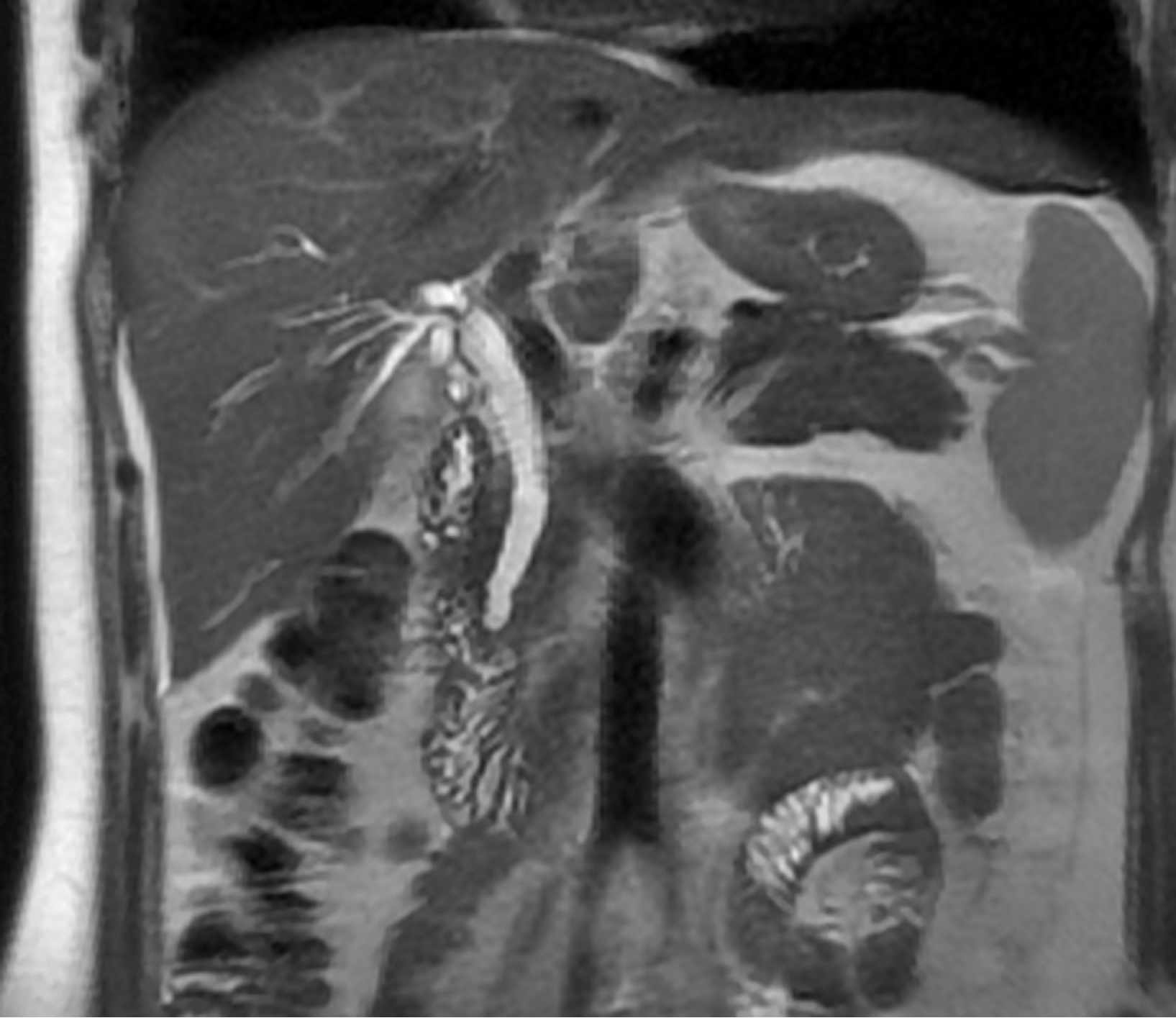
Figure 1
Magnetic resonance cholangiopancreatography showed that the stone was located in the sphincter of Oddi at the terminal common bile duct.
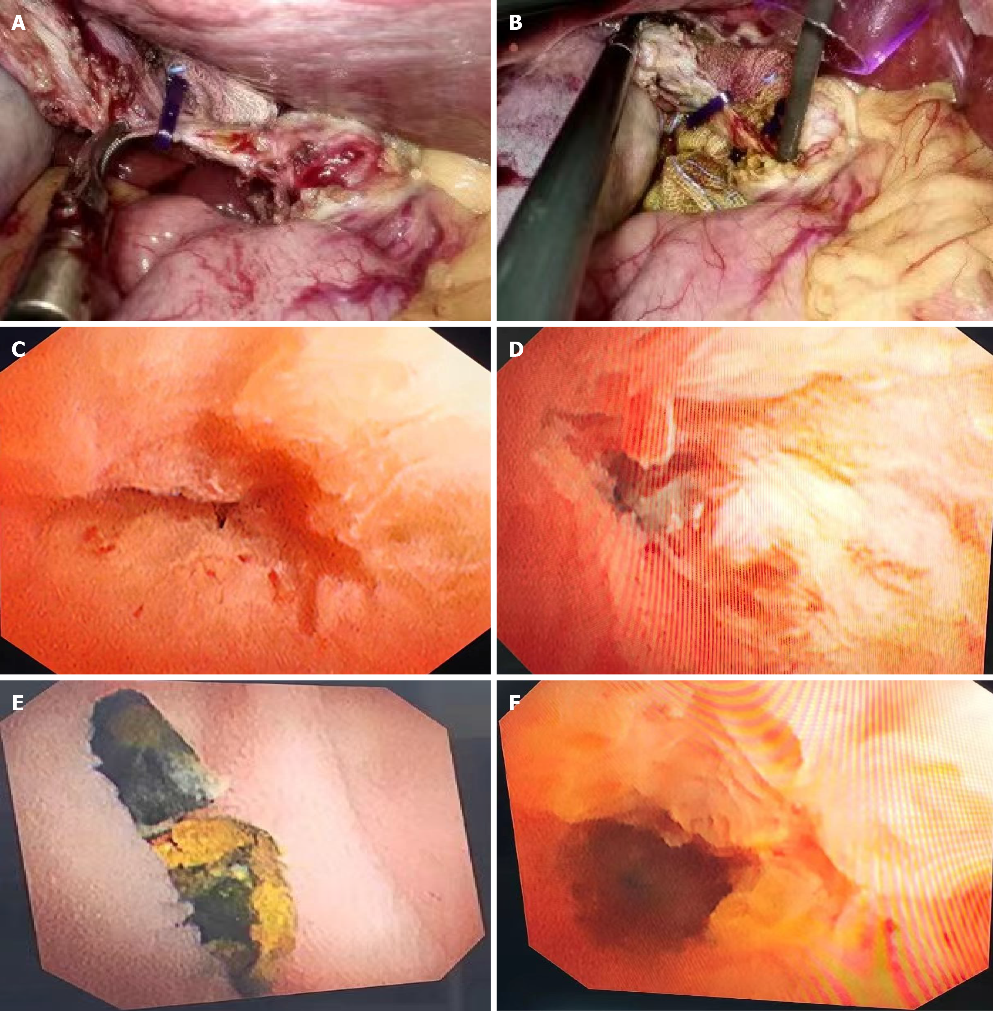
Figure 2 The process of Oddi intersphincter stone removal by ultrafine choledochoscopy combined with low-dose atropine through the gallbladder duct.
A: After retrograde cholecystectomy, the anterior wall of the gallbladder duct was cut longitudinal at 0.5 mm from the common bile duct; B: Ultrafine choledochoscopy was inserted through the gallbladder duct to explore the common bile duct; C: Routine exploration of common bile duct to the terminal sphincter of Oddi showed no residual stone; D: The ultrafine choledochoscopy entered into the sphincter of Oddi, and the stones were embedded in the lower zone; E: By using atropine, the stones were pushed into the intestinal cavity; F: The surgical field of relaxation of Oddi sphincter after stone removal.
- Citation: Hu XS, Wang Y, Pan HT, Zhu C, Zhou S, Chen SL, Liu HC, Pang Q, Jin H. Initial experience with ultrafine choledochoscopy combined with low-dose atropine for the treatment of Oddi intersphincter stones. World J Gastrointest Surg 2025; 17(3): 102998
- URL: https://www.wjgnet.com/1948-9366/full/v17/i3/102998.htm
- DOI: https://dx.doi.org/10.4240/wjgs.v17.i3.102998


