©The Author(s) 2025.
World J Gastrointest Surg. Oct 27, 2025; 17(10): 111004
Published online Oct 27, 2025. doi: 10.4240/wjgs.v17.i10.111004
Published online Oct 27, 2025. doi: 10.4240/wjgs.v17.i10.111004
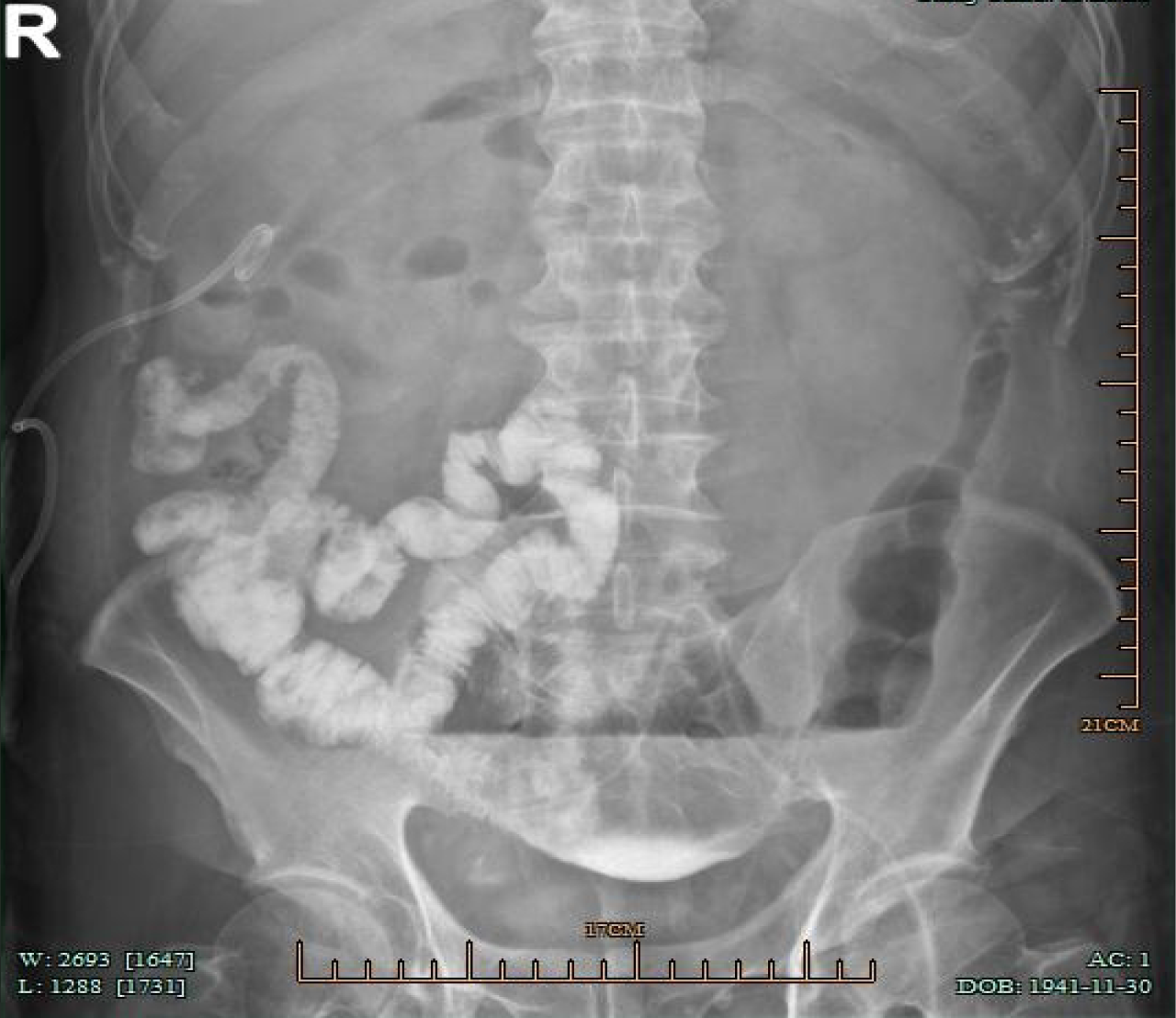
Figure 1
After injection of contrast agent through the original abdominal drainage tube in the left lower abdomen, the abdominal plain radiograph showed the small intestine in the right middle and lower abdomen filled with contrast agent.
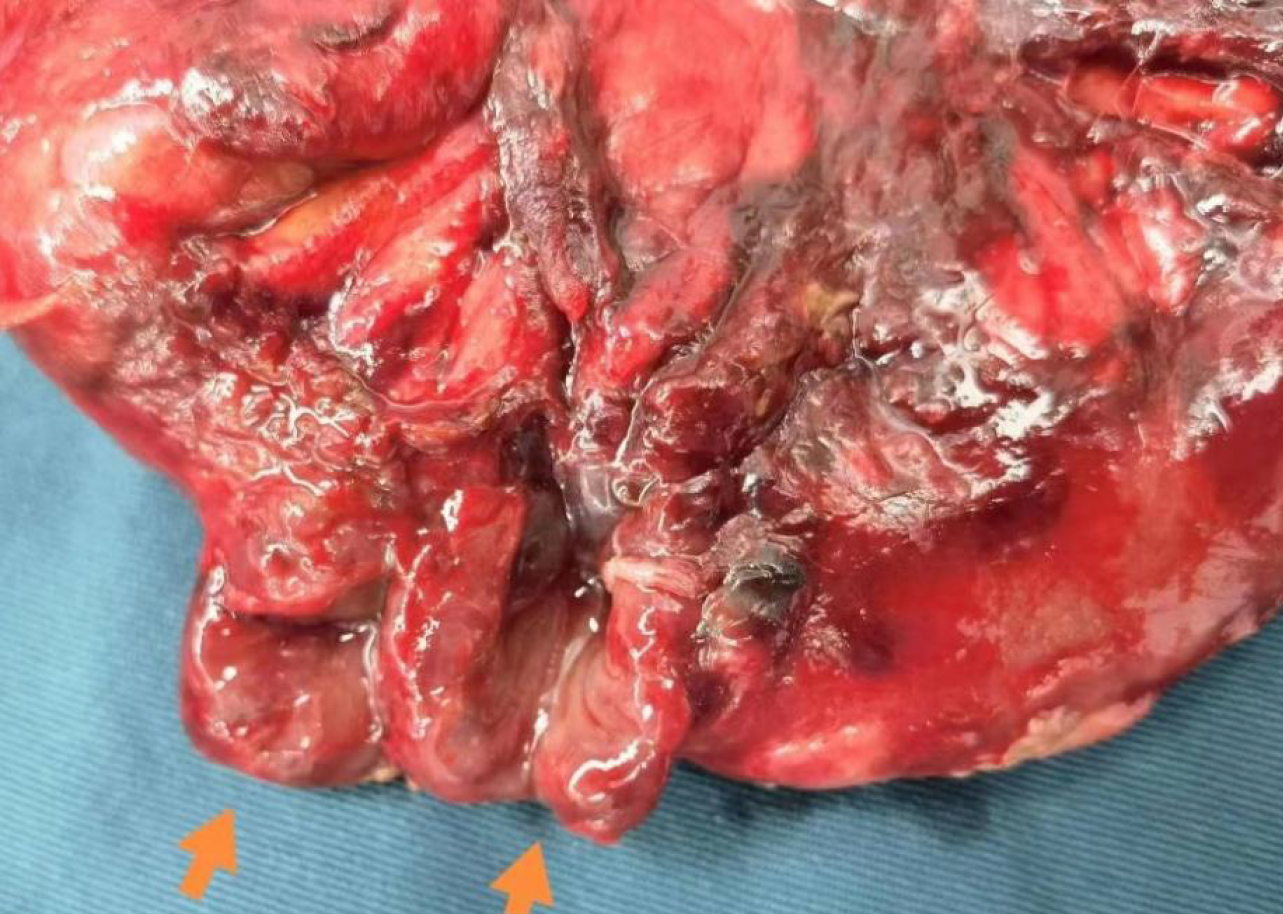
Figure 2 Two adjacent small intestine ruptures with a diameter of approximately 1.
5 cm were observed during the operation. The rupture sites had overflow of small intestinal fluid, with surrounding granulation tissue encapsulation.
- Citation: Pan XF, Cai Y, Cao YG. Small intestinal fistula caused by drainage tube removal: A case report. World J Gastrointest Surg 2025; 17(10): 111004
- URL: https://www.wjgnet.com/1948-9366/full/v17/i10/111004.htm
- DOI: https://dx.doi.org/10.4240/wjgs.v17.i10.111004


