Copyright
©The Author(s) 2024.
World J Gastrointest Surg. Aug 27, 2024; 16(8): 2649-2661
Published online Aug 27, 2024. doi: 10.4240/wjgs.v16.i8.2649
Published online Aug 27, 2024. doi: 10.4240/wjgs.v16.i8.2649
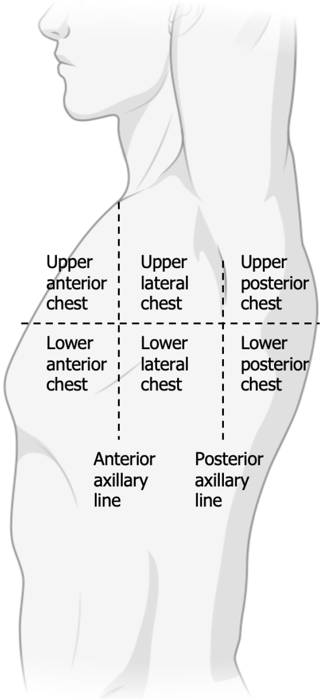
Figure 1 Locations of pulmonary ultrasound being recorded on one side.
The anterior, lateral, and posterior chest are separated by the anterior/posterior axillary lines.
Figure 2 The pulmonary ultrasound images for different scores.
A: 0 points, 0 to 2 B lines; B: 1 point, 3 or more B lines, thickened pleural line; C: 2 points, multiple overlapped B lines, thickened or irregular pleural line.
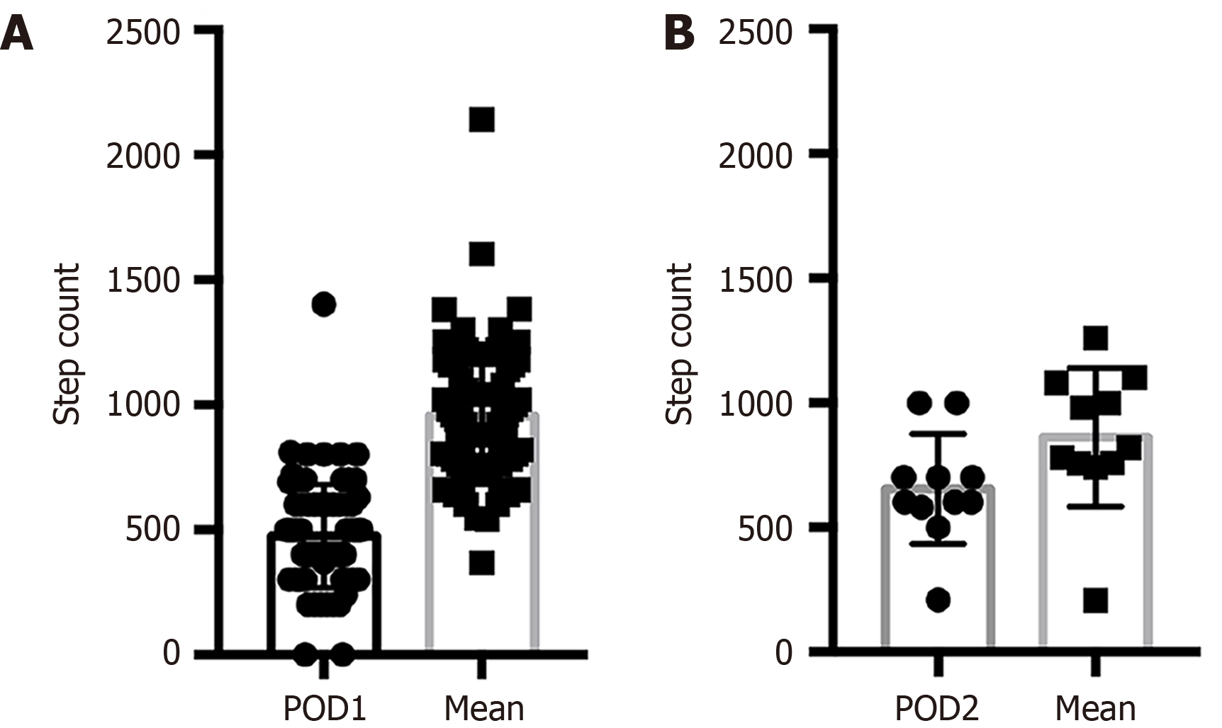
Figure 3 Comparison of step counts in patients with the largest increase of pulmonary ultrasound between different postoperative days and mean step counts of all patients.
A: Patients with the largest increase in pulmonary ultrasound score between postoperative day (POD) 1 and POD2, n = 79, P < 0.05; B: Patients with the largest increase in pulmonary ultrasound score between POD2 and POD3, n = 11, P < 0.05. POD: Postoperative day.
- Citation: Lin C, Wang PP, Wang ZY, Lan GR, Xu KW, Yu CH, Wu B. Innovative integration of lung ultrasound and wearable monitoring for predicting pulmonary complications in colorectal surgery: A prospective study. World J Gastrointest Surg 2024; 16(8): 2649-2661
- URL: https://www.wjgnet.com/1948-9366/full/v16/i8/2649.htm
- DOI: https://dx.doi.org/10.4240/wjgs.v16.i8.2649


