Copyright
©The Author(s) 2024.
World J Gastrointest Surg. Jul 27, 2024; 16(7): 2329-2336
Published online Jul 27, 2024. doi: 10.4240/wjgs.v16.i7.2329
Published online Jul 27, 2024. doi: 10.4240/wjgs.v16.i7.2329
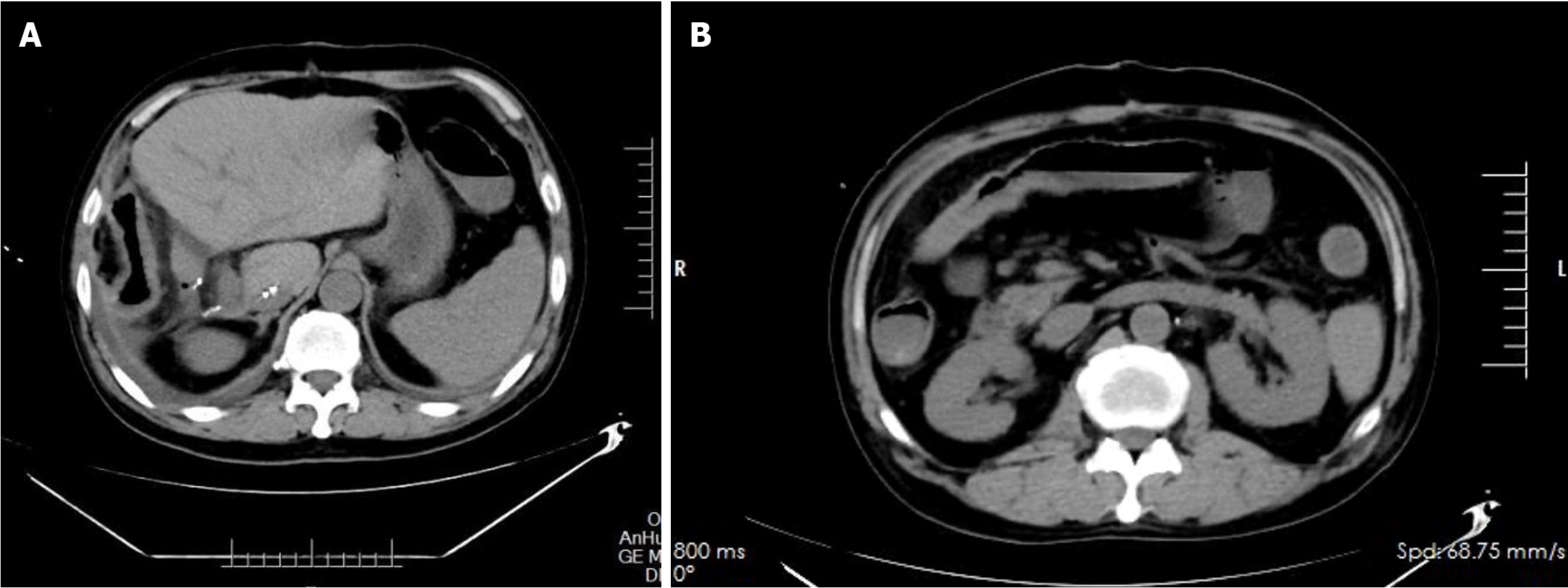
Figure 1 Full abdominal pelvic spiral computed tomography plain scan on August 20, 2022.
A: The wall of the ascending colon was slightly thickened and showed uniform edema, while the surrounding fat space was slightly blurred; B: The wall of the transverse colon was slightly thickened, and moderate lymph nodes were scattered. There was no obvious lesion in the small intestine.
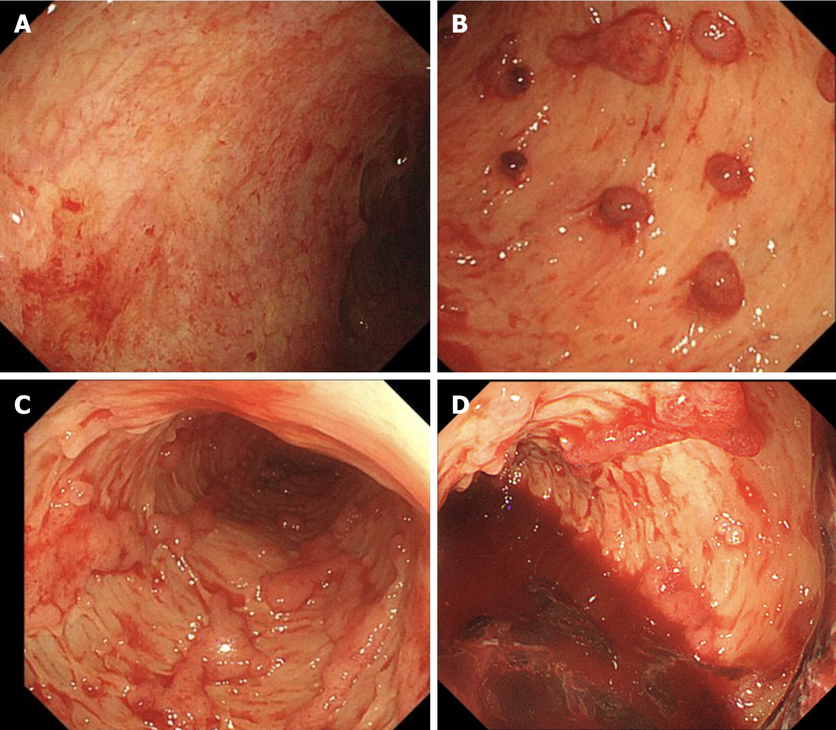
Figure 2 Sigmoidoscopy examination.
A: The lesions in the rectum, which were less than 5 cm, appeared to be relatively mild; B and C: Extensive mucosal exfoliation was observed below 30 cm in the sigmoid colon, along with scattered, small, dotted mucous membranes; D: A significant presence of blood and clots was found in the sigmoid colon.
Figure 3 Pathology of the sigmoidoscopic biopsy specimen.
A: Ulcer formation: Surface inflammatory exudation necrosis, underlying granulation tissue hyperplasia, interstitial "transmural" hybrid inflammatory cell infiltration, hematoxylin and eosin (HE) staining × 40; B: Cryptitis, crypt abscesses (yellow arrow), and significantly increased numbers of apoptotic cells (blue arrows), HE × 400.
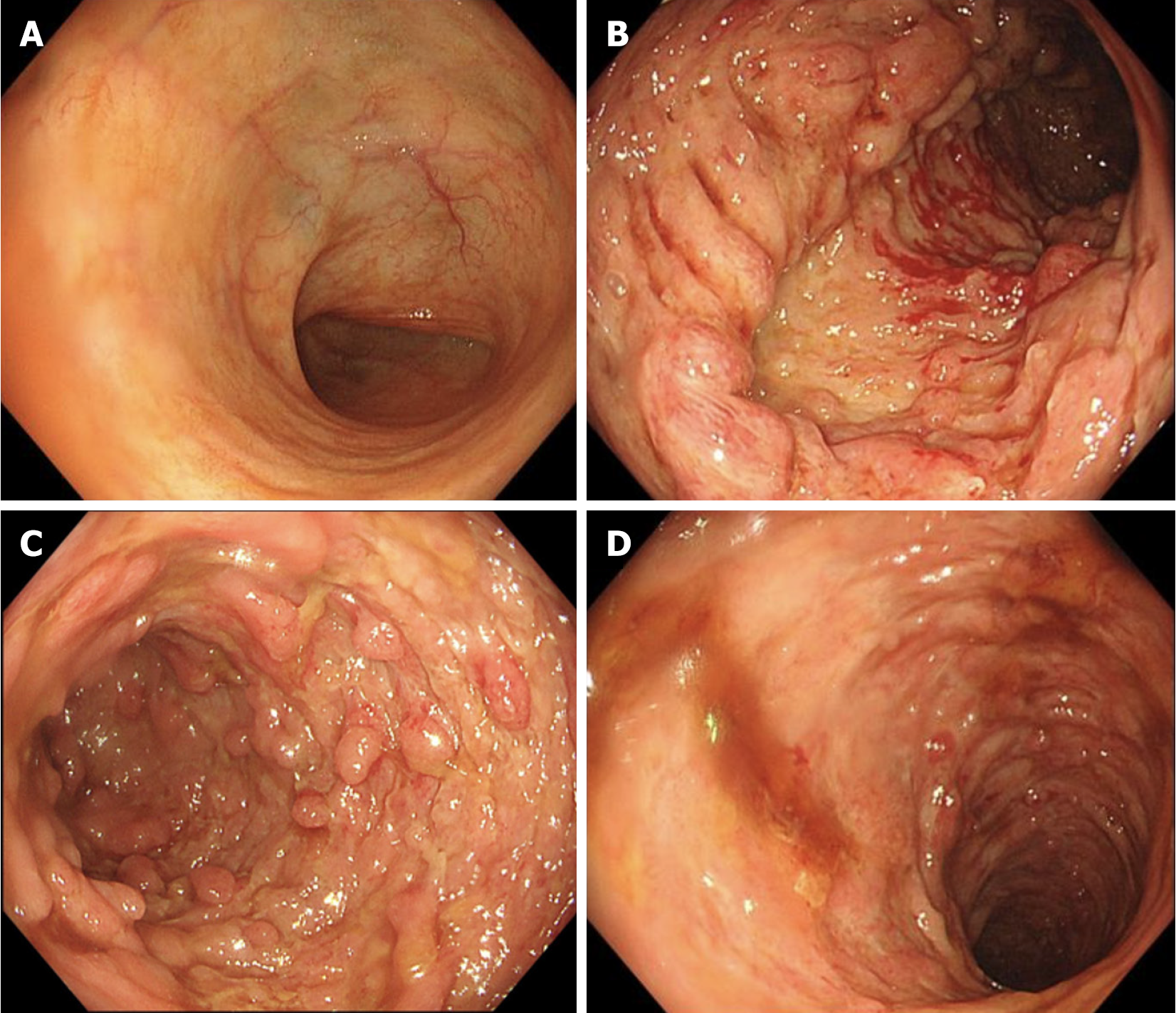
Figure 4 Colonoscopy examination.
A: The mucosa of the terminal ileum is normal; B and C: Extensive and continuous hyperemia, edema, erosion, polypoid hyperplasia, irregular deep chisel and geoglyphic ulcers; D: The ulcers and erosion of the sigmoid colon were significantly improved.
Figure 5 Postoperative pathology.
A: Ulcer formation, surface bleeding with inflammatory exudation, necrosis, mucous membrane accompanied by hemorrhagic changes. Hematoxylin and eosin (HE) staining × 40; B: Apoptotic cells were found in the crypt (blue arrow). HE × 400.
- Citation: Hong N, Wang B, Zhou HC, Wu ZX, Fang HY, Song GQ, Yu Y. Multidisciplinary management of ulcerative colitis complicated by immune checkpoint inhibitor-associated colitis with life-threatening gastrointestinal hemorrhage: A case report. World J Gastrointest Surg 2024; 16(7): 2329-2336
- URL: https://www.wjgnet.com/1948-9366/full/v16/i7/2329.htm
- DOI: https://dx.doi.org/10.4240/wjgs.v16.i7.2329


