©The Author(s) 2024.
World J Gastrointest Surg. May 27, 2024; 16(5): 1461-1466
Published online May 27, 2024. doi: 10.4240/wjgs.v16.i5.1461
Published online May 27, 2024. doi: 10.4240/wjgs.v16.i5.1461
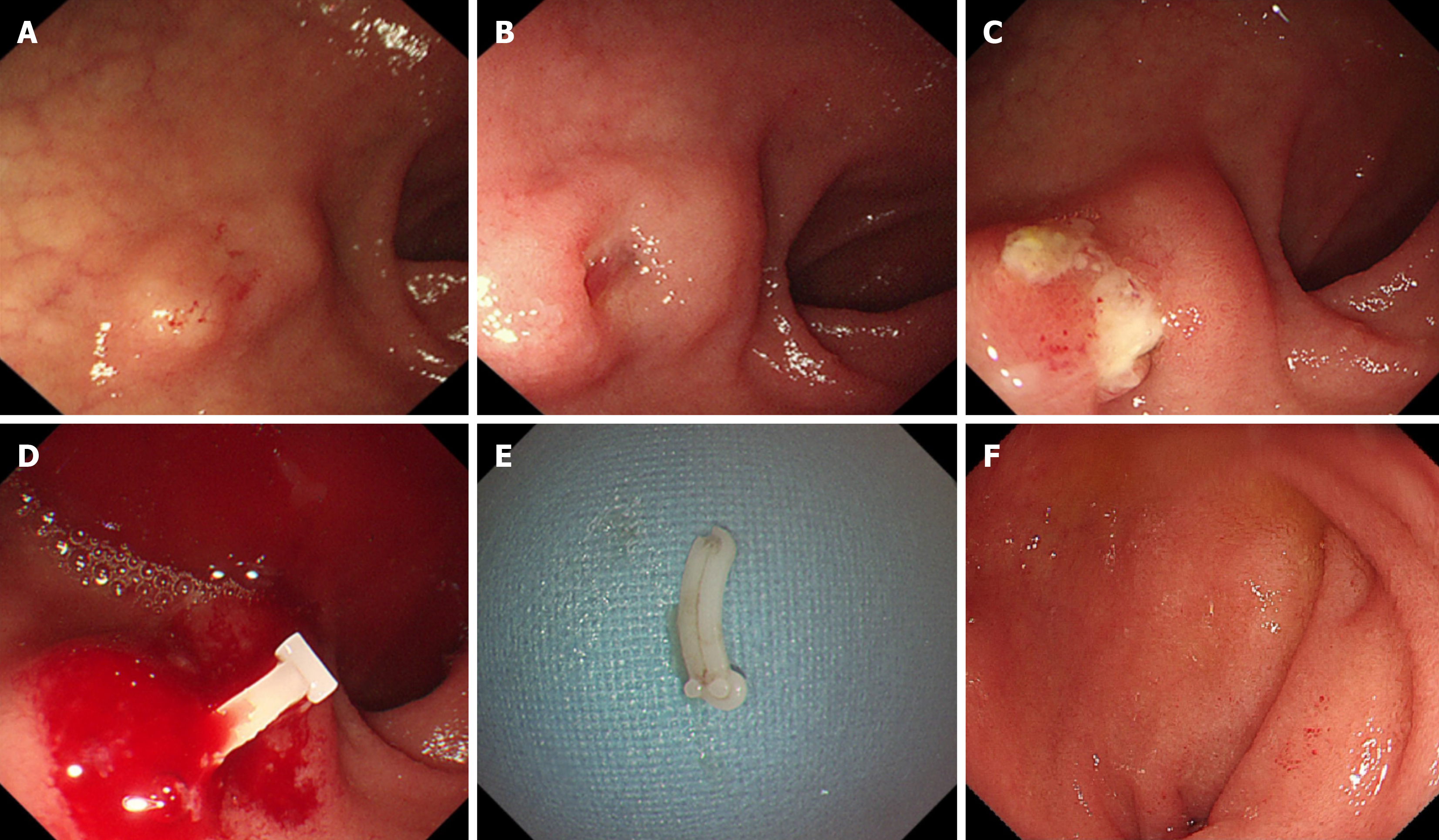
Figure 1 The esophagogastroduodenoscopy results.
A: The first esophagogastroduodenoscopy (EGD) revealed a submucosal tumor (SMT)-like lesion in the duodenal bulb with normal overlying mucosa; B: The second EGD revealed an SMT-like lesion in the duodenal bulb with raised edges and central depression; C: The third EGD revealed an SMT-like lesion, covered by white exudates, with erosions and edema. The lesion was active and hard when touched with biopsy forceps; D: Olympus grasping forceps removed the foreign body; E: The foreign body was a Hem-o-lok clip; F: The fourth EGD revealed the duodenum was covered with normal mucosa.
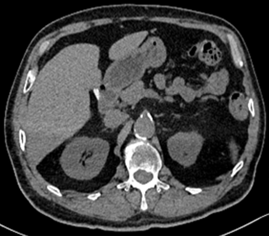
Figure 2 Computed tomography scan examination.
Computed tomography scan shows a foreign body in the duodenum.
- Citation: Liu HY, Yin AH, Wei Z. Hem-o-lok clip migration to duodenal bulb post-cholecystectomy: A case report. World J Gastrointest Surg 2024; 16(5): 1461-1466
- URL: https://www.wjgnet.com/1948-9366/full/v16/i5/1461.htm
- DOI: https://dx.doi.org/10.4240/wjgs.v16.i5.1461


