©The Author(s) 2024.
World J Gastrointest Surg. May 27, 2024; 16(5): 1436-1442
Published online May 27, 2024. doi: 10.4240/wjgs.v16.i5.1436
Published online May 27, 2024. doi: 10.4240/wjgs.v16.i5.1436
Figure 1 Timeline of history of past illnessthe, initial diagnosis, surgical intervention, postoperative adjuvant therapy, and follow-up period.
PET-CT: Positron emission tomography/computed tomography; PD-1: programmed cell death 1.
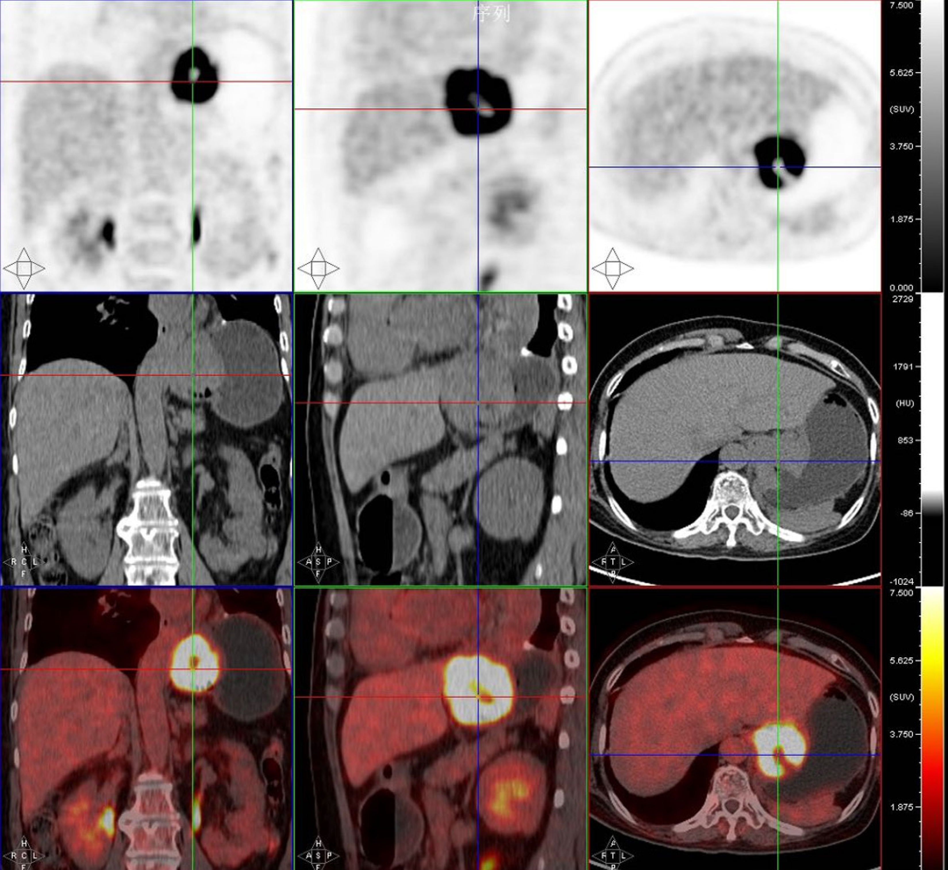
Figure 2 Positron emission tomography/computed tomography showing high accumulation of 18F-fludeoxyglucose in the gastric cardia region.
A maximum standardized uptake value of 20.03.
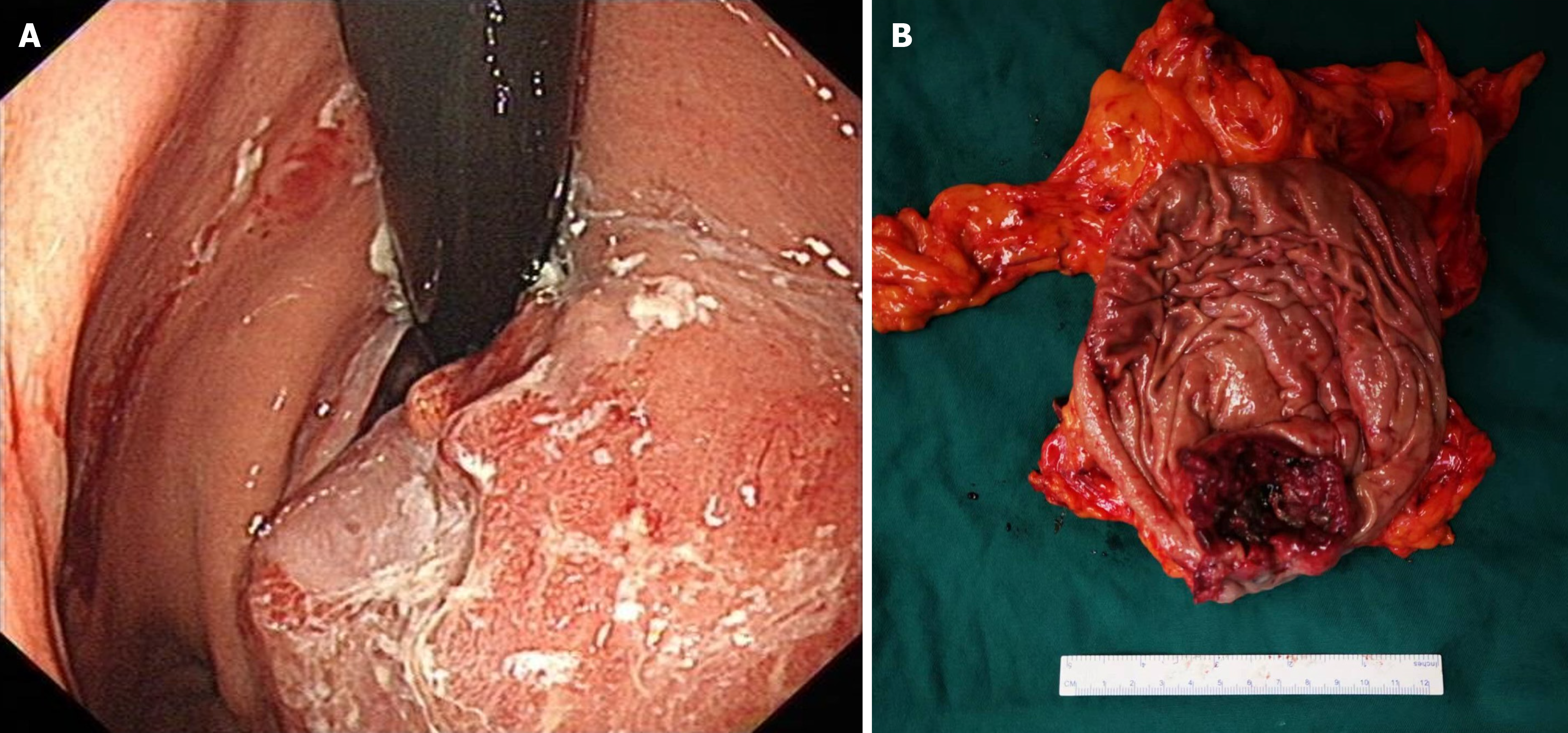
Figure 3 Upper endoscopy and surgical resection.
A: The presence of a large mass of about 5 cm × 4 cm at the stomach fundus was confirmed by upper endoscopy; B: Surgical resection of the tumor revealed a large mass located in the stomach fundus near the cardia, showing a type of ulcer infiltrate measuring 6 cm × 5 cm.
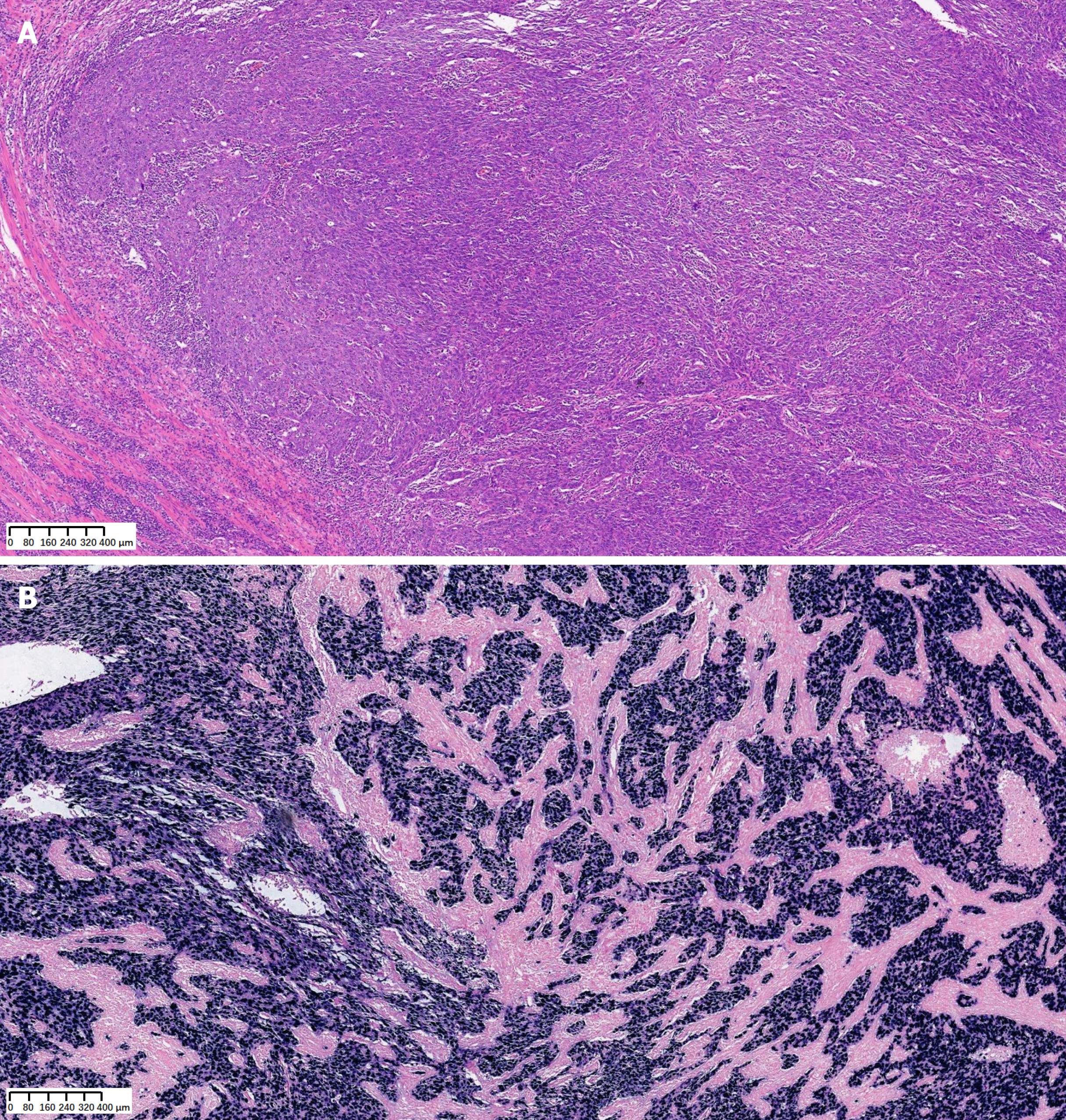
Figure 4 Hematoxylin and eosin staining and in situ hybridization.
A: Histopathological examination of a stomach tumor section by hematoxylin and eosin staining, showing poorly differentiated carcinoma with prominent lymphoplasmacytic infiltration; B: In situ hybridization analysis showing positive staining for Epstein-Barr virus-encoded RNA.
- Citation: Chen GF, Wang J, Yan Y, Xu S, Chen J. Metastatic stomach lymphoepithelioma-like carcinoma and immune checkpoint inhibitor therapy: A case report. World J Gastrointest Surg 2024; 16(5): 1436-1442
- URL: https://www.wjgnet.com/1948-9366/full/v16/i5/1436.htm
- DOI: https://dx.doi.org/10.4240/wjgs.v16.i5.1436


