©The Author(s) 2024.
World J Gastrointest Surg. Apr 27, 2024; 16(4): 1208-1214
Published online Apr 27, 2024. doi: 10.4240/wjgs.v16.i4.1208
Published online Apr 27, 2024. doi: 10.4240/wjgs.v16.i4.1208
Figure 1 Different sections of abdominal contrast-enhanced computed tomography image showing nonenhancing masses on diseased jejunal mesentery (orange arrow).
A: Sagittal plane image; B: Coronal plane image; C: Transverse plane image.
Figure 2 Representative images of single-balloon enteroscopy.
A: Characteristic endoscopic manifestations of edematous white-yellow mucosa accompanied by hemorrhagic red spots (white arrow); B and C: Images demonstrate multiple active bleeding lesions (white arrow).
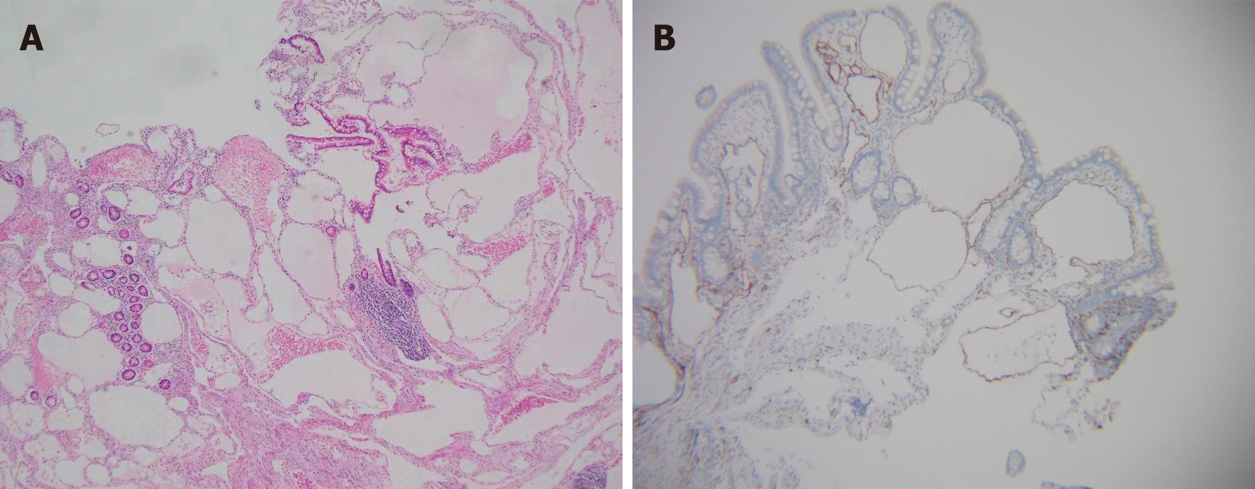
Figure 3 Microscopic features of the biopsy specimens.
A: The mucosal lamina propria and submucosa exhibited variably sized cysts, which were filled with lymphatic fluid and contained red blood cells and lymphocytes within the lumen (hematoxylin–eosin staining); B: Positive immunohistochemical staining for D2-40 was observed.
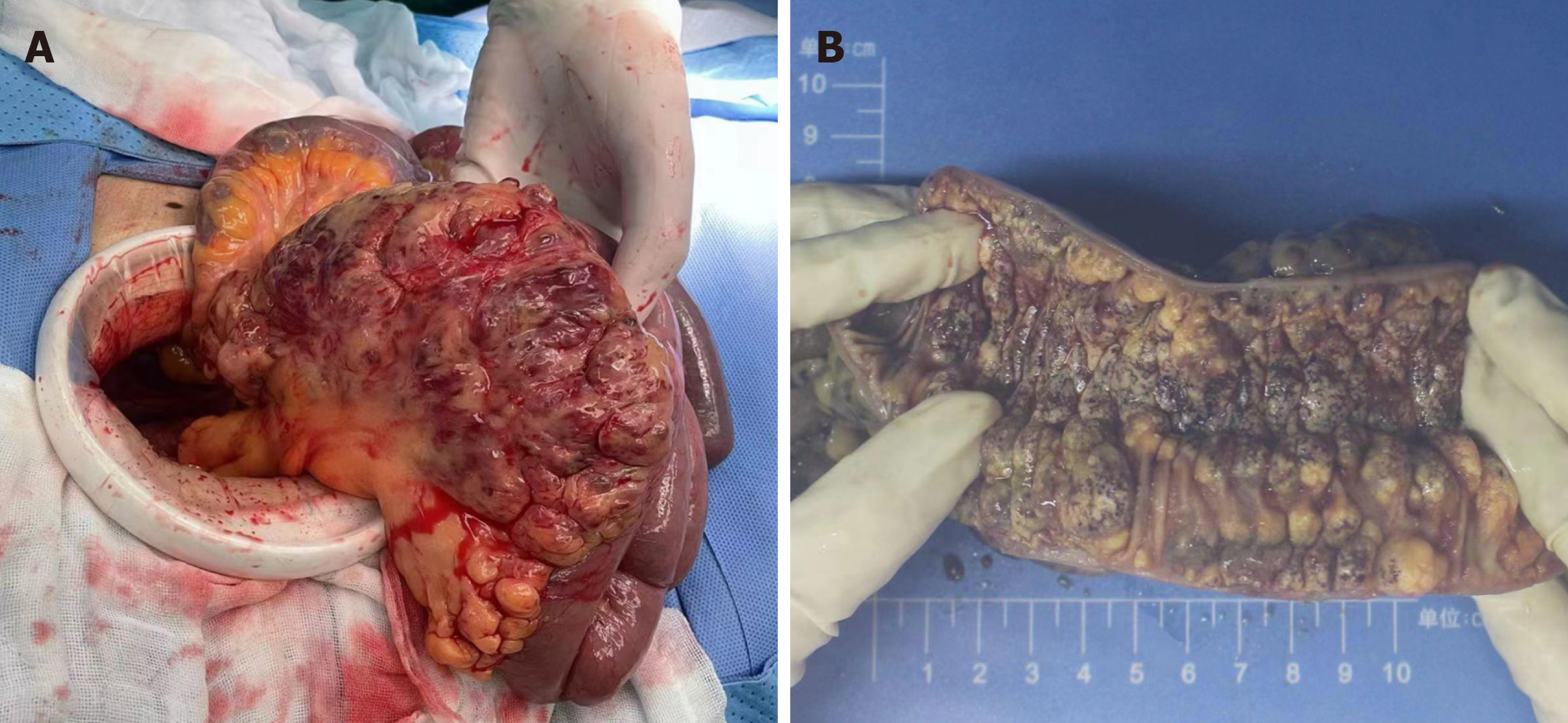
Figure 4 Intraoperative findings and resection specimen.
A: Numerous cystic and hemorrhagic masses of varying sizes were observed on the mesentery; B: The length of the diseased jejunum exceeded 10 cm. Longitudinal gross dissected specimens revealed that various sizes of nodular eminence were distributed on the lumen surface.
- Citation: Liu KR, Zhang S, Chen WR, Huang YX, Li XG. Intermittent melena and refractory anemia due to jejunal cavernous lymphangioma: A case report. World J Gastrointest Surg 2024; 16(4): 1208-1214
- URL: https://www.wjgnet.com/1948-9366/full/v16/i4/1208.htm
- DOI: https://dx.doi.org/10.4240/wjgs.v16.i4.1208


