©The Author(s) 2024.
World J Gastrointest Surg. Nov 27, 2024; 16(11): 3471-3483
Published online Nov 27, 2024. doi: 10.4240/wjgs.v16.i11.3471
Published online Nov 27, 2024. doi: 10.4240/wjgs.v16.i11.3471
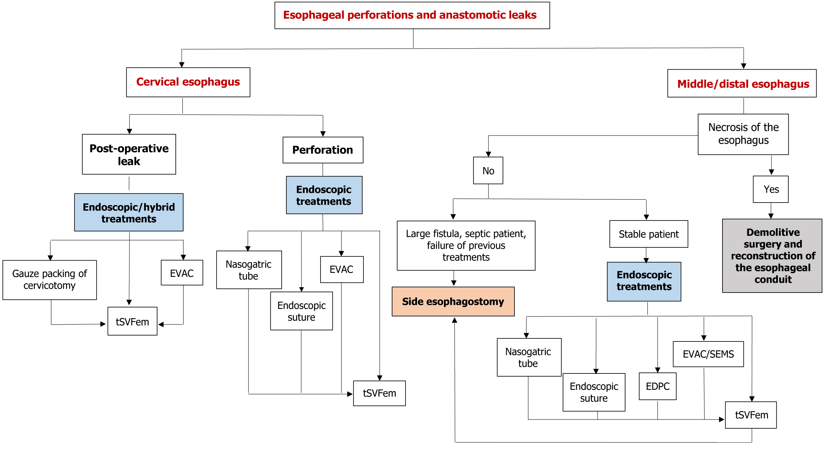
Figure 1 Flowchart summarizing our multidisciplinary endoscopic, hybrid (endoscopic + surgical) or surgical management of esophageal perforations and postoperative leaks.
EDPC: Endoscopic double-pigtail catheter; EVAC: Endoscopic vacuum-assisted closure therapy; SEMS: Self-expanding-metal stent; tSVFem: Emulsified autologous stromal vascular fraction.
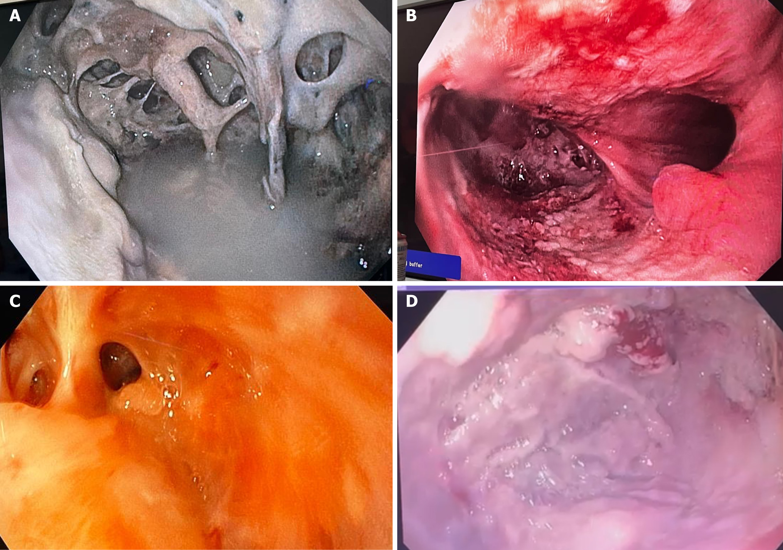
Figure 2 Endoscopic image of anastomotic leak after distal esophagus diverticulatectomy.
A: Initial aspect; B and C: After consecutive endoscopic vacuum-assisted closure therapy treatment; D: Complete resolution.
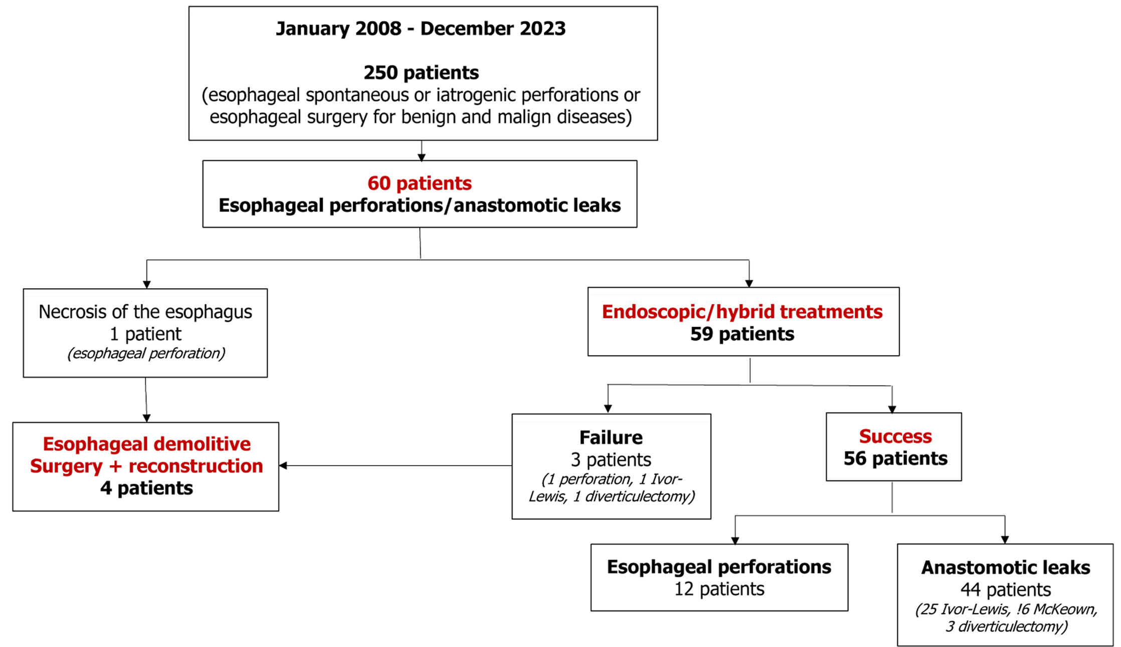
Figure 3 Management of the whole population of the study.
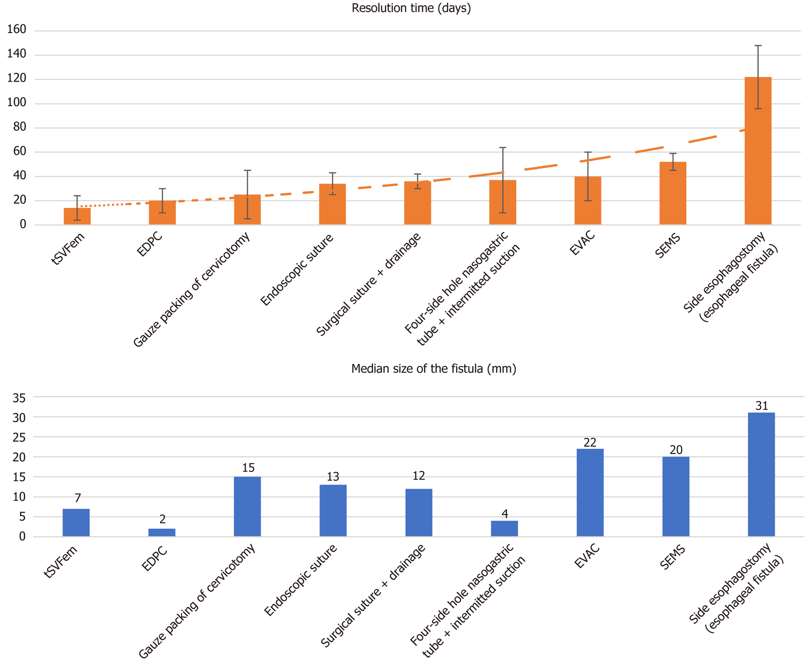
Figure 4 Resolution time and corresponding median diameter of the esophageal fistula per each type of treatment adopted in the study.
EDPC: Endoscopic double-pigtail catheter; EVAC: Endoscopic vacuum-assisted closure therapy; SEMS: Self-expanding-metal stent; tSVFem: Emulsified autologous stromal vascular fraction.
- Citation: Nachira D, Calabrese G, Senatore A, Pontecorvi V, Kuzmych K, Belletatti C, Boskoski I, Meacci E, Biondi A, Raveglia F, Bove V, Congedo MT, Vita ML, Santoro G, Petracca Ciavarella L, Lococo F, Punzo G, Trivisonno A, Petrella F, Barbaro F, Spada C, D'Ugo D, Cioffi U, Margaritora S. How to preserve the native or reconstructed esophagus after perforations or postoperative leaks: A multidisciplinary 15-year experience. World J Gastrointest Surg 2024; 16(11): 3471-3483
- URL: https://www.wjgnet.com/1948-9366/full/v16/i11/3471.htm
- DOI: https://dx.doi.org/10.4240/wjgs.v16.i11.3471


