©The Author(s) 2024.
World J Gastrointest Surg. Nov 27, 2024; 16(11): 3425-3436
Published online Nov 27, 2024. doi: 10.4240/wjgs.v16.i11.3425
Published online Nov 27, 2024. doi: 10.4240/wjgs.v16.i11.3425
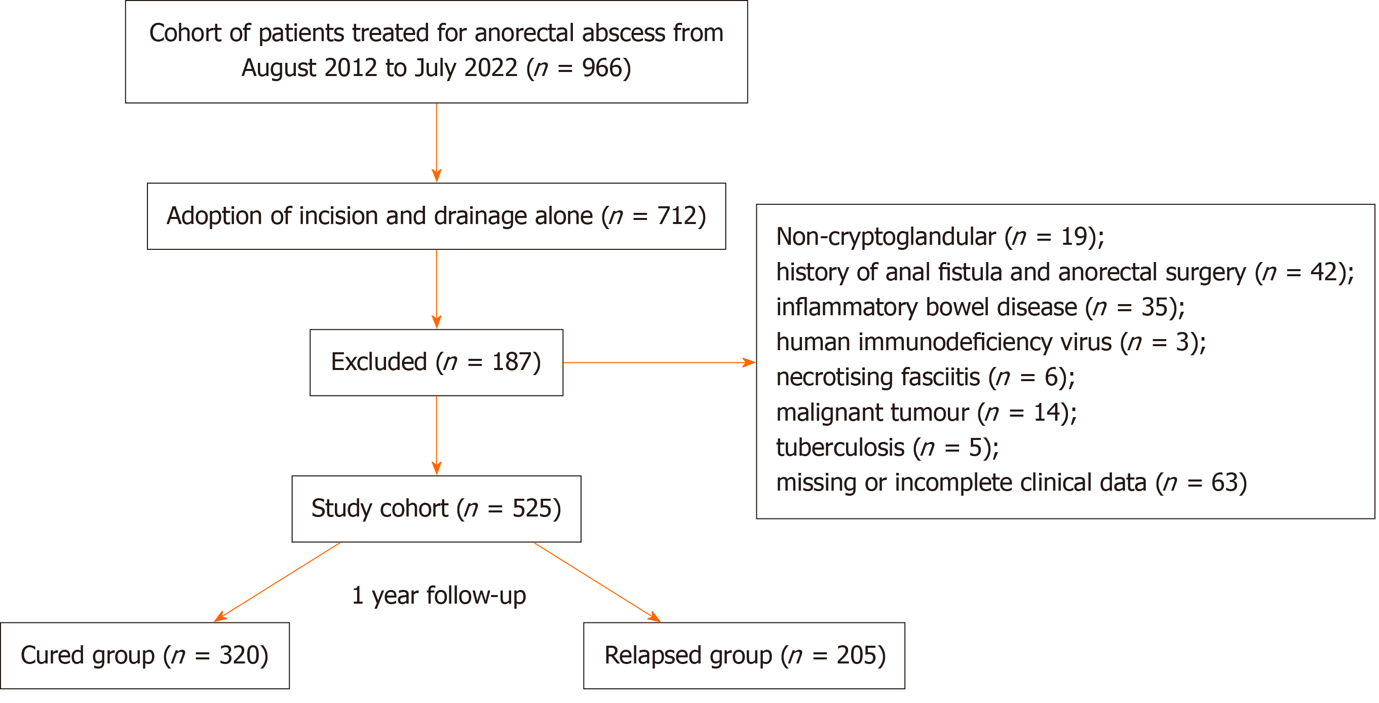
Figure 1 The flowchart of study design.
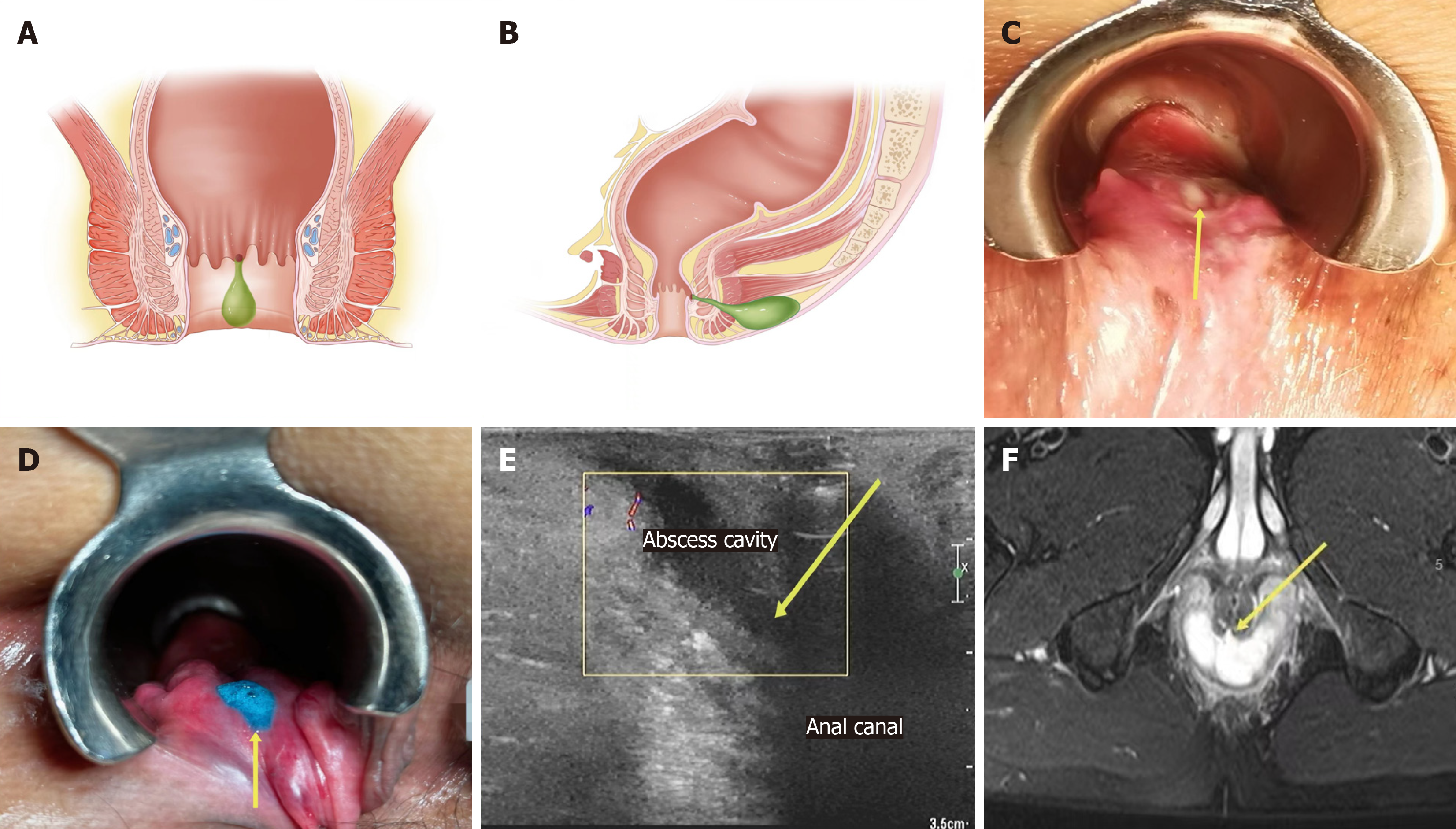
Figure 2 Clinical characteristics of fistula-prone abscess.
A: There is a hard nodule, a depression, even a defect at the dentate line; B: Sclerotic tissue between the pus cavity and the anal canal by a finger touch; C: Pus oozes out of the corresponding area when press the pus cavity (direction of the yellow arrow); D: Liquid overflows when instilling hydrogen peroxide + methylene blue into the pus cavity during the operation (direction of the yellow arrow); E: Abscess cavity leading to the anal canal, internal sphincter involvement or discontinuity (direction of the yellow arrow in the ultrasound imaging); F: Abscess cavity leading to the anal canal, internal sphincter involvement or discontinuity (direction of the yellow arrow in the magnetic resonance imaging).
- Citation: Chen SZ, Sun KJ, Gu YF, Zhao HY, Wang D, Shi YF, Shi RJ. Proposal for a new classification of anorectal abscesses based on clinical characteristics and postoperative recurrence. World J Gastrointest Surg 2024; 16(11): 3425-3436
- URL: https://www.wjgnet.com/1948-9366/full/v16/i11/3425.htm
- DOI: https://dx.doi.org/10.4240/wjgs.v16.i11.3425


