Copyright
©The Author(s) 2023.
World J Gastrointest Surg. Aug 27, 2023; 15(8): 1574-1590
Published online Aug 27, 2023. doi: 10.4240/wjgs.v15.i8.1574
Published online Aug 27, 2023. doi: 10.4240/wjgs.v15.i8.1574
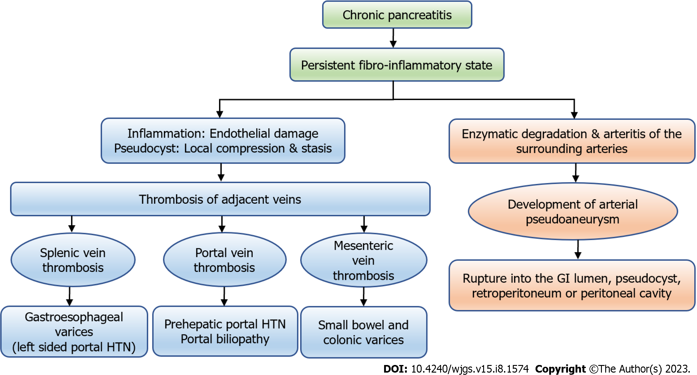
Figure 1 The types of vascular complications in chronic pancreatitis and their consequences.
GI: Gastrointestinal; HTN: Hypertension.
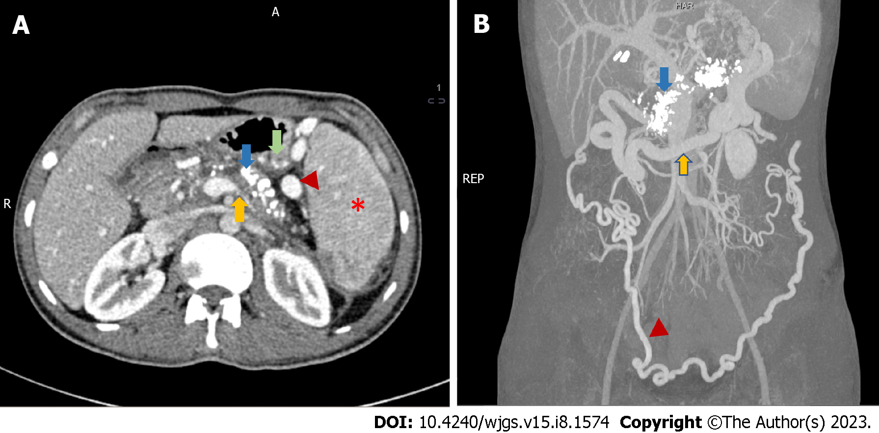
Figure 2 Radiological features of chronic calcific pancreatitis and its complications including venous thrombosis and collaterals.
A: An axial section of contrast-enhanced computed tomography (CECT) showing chronic pancreatitis with calcifications (blue arrow), attenuated splenic vein (yellow arrow), multiple perigastric collaterals (green arrow), gastrosplenic collaterals (red arrowhead) and splenomegaly (red asterisk); B: Coronal section of CECT of the same patient showing extensive pancreatic calcification (blue arrow) with dilated gastroepiploic vein (yellow arrow) and omental collaterals (red arrowhead). Courtesy: Dr Madhusudhan KS, Department of Radiodiagnosis.
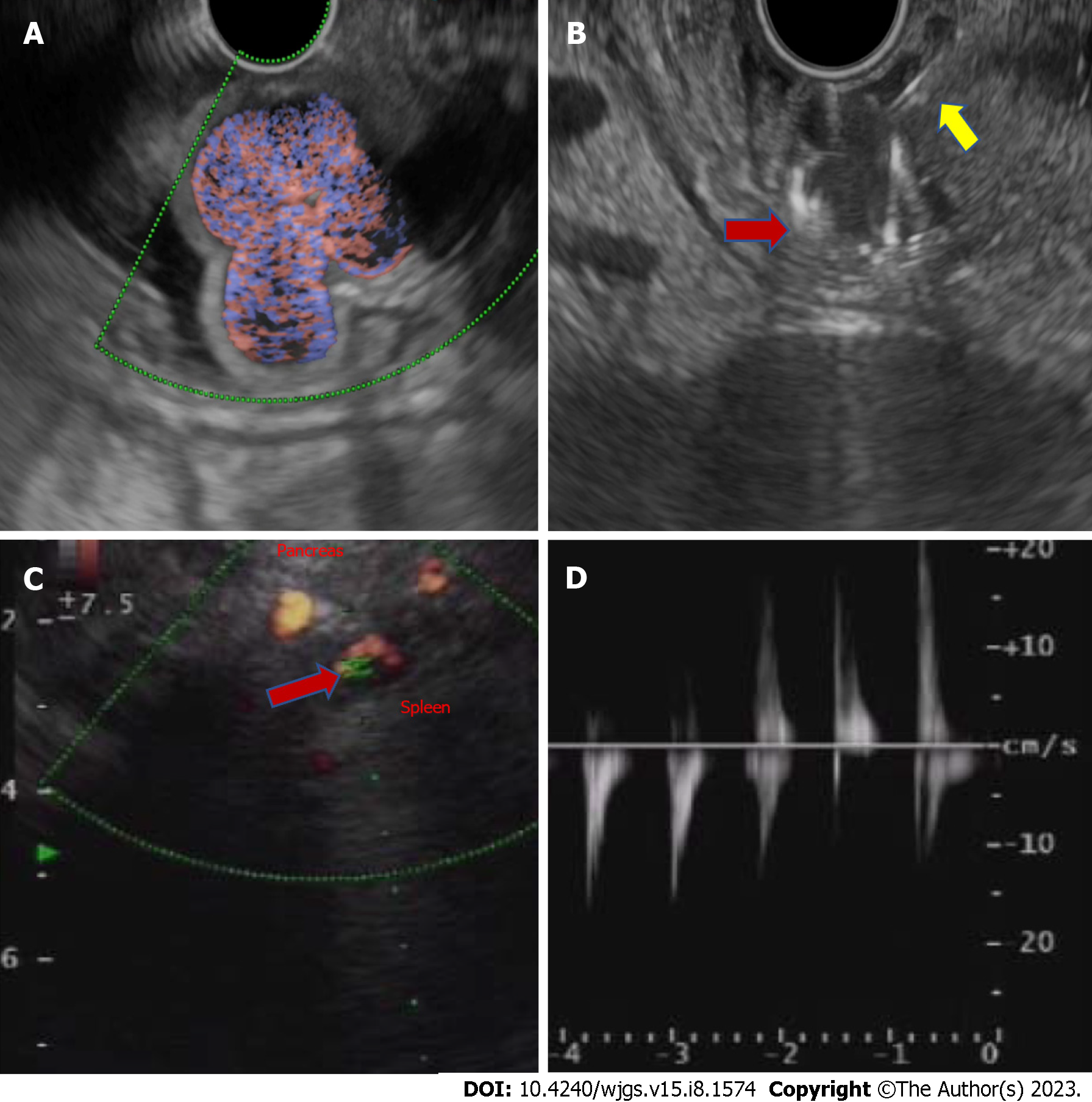
Figure 3 Endoscopic ultrasound-guided fundal variceal obliteration, and pseudoaneurysm on endoscopic ultrasound.
A: Linear endoscopic ultrasound showing the fundal varices on doppler study; B: Linear endoscopic ultrasound guided metal coil (red arrow, hyperechoic curved structure) being pushed into the varices for obliteration after the endoscopic ultrasound needle puncture (yellow arrow, hyperechoic linear structure); C: Linear endoscopic ultrasound showing an arterial pseudoaneurysm (red arrow) on doppler study; D: On power doppler mode Doppler showing an arterial waveform with bidirectional flow, classically labelled as “yin-yang” sign.
Figure 4 Arterial pseudoaneurysm in chronic pancreatitis.
A: Axial contrast-enhanced computed tomography in a patient of chronic pancreatitis showing a pseudoaneurysm (PsA) (red arrow) arising from splenic artery along with a specks of parenchymal calcification (green arrow) and a pseudocyst (yellow arrow) in the head of pancreas; B: Reconstructed angiographic image of the same patient showing splenic artery PsA (white arrow); C: Digital subtraction angiography of the same patient showing contrast outpouching from the splenic artery suggestive of splenic artery PsA (red arrow) with contrast opacification before endovascular therapy. Courtesy: Dr Madhusudhan KS, Department of Radiodiagnosis.
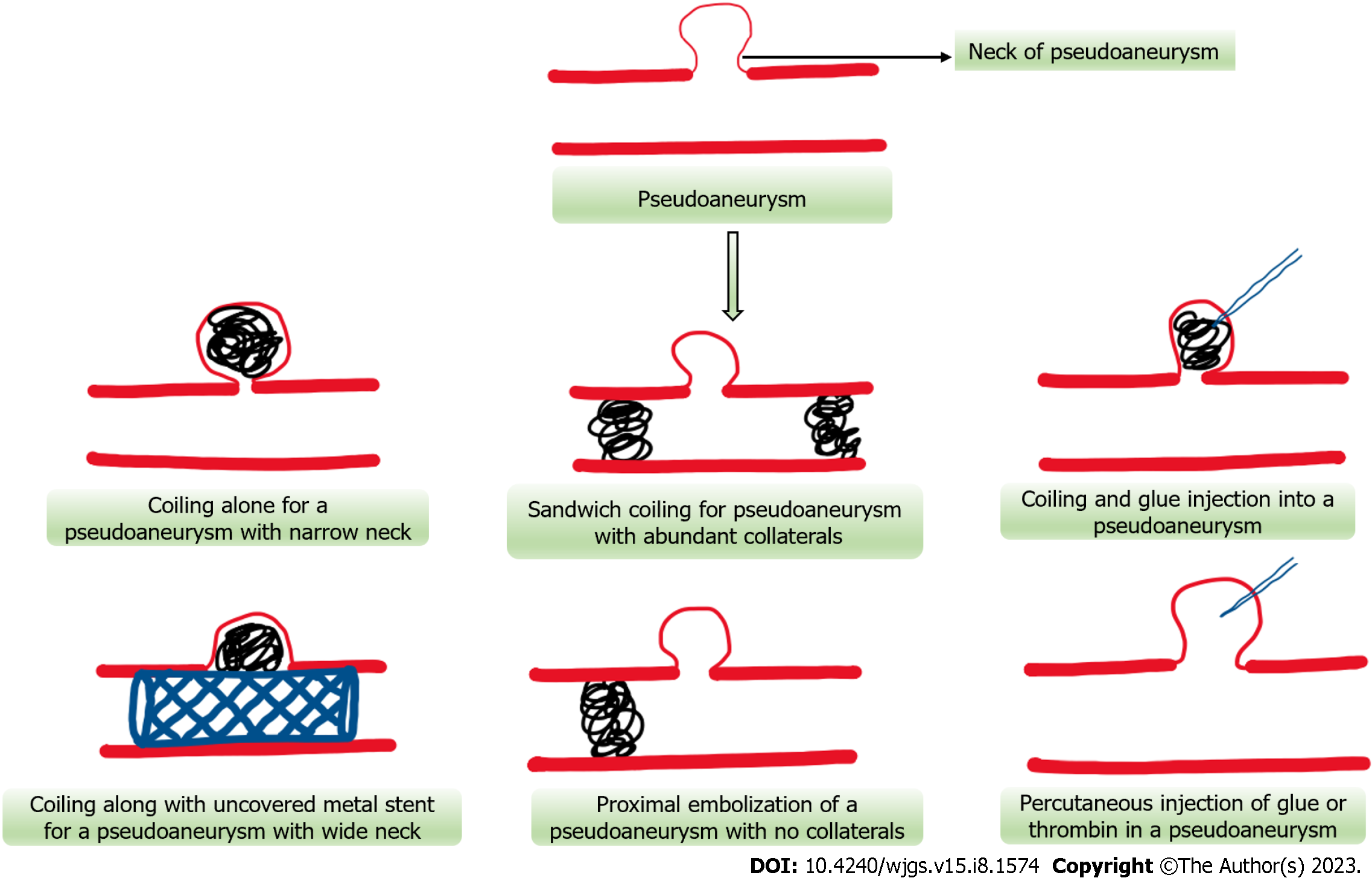
Figure 5 Schematic representation of various endovascular and percutaneous approaches for pseudoaneurysm obliteration.
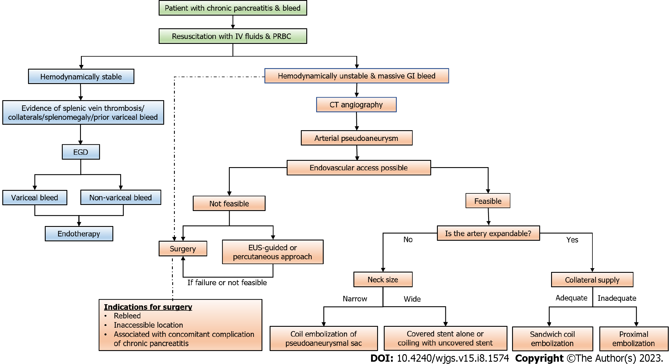
Figure 6 Flow diagram depicting the approach to a case of gastrointestinal bleeding in patients with chronic pancreatitis (dashed arrow suggests an alternative management strategy).
CT: Computed tomography; EGD: Esophagogastroduodenoscopy; GI: Gastrointestinal; PRBC: Packed red blood cell; EUS: Endoscopic ultrasound.
- Citation: Walia D, Saraya A, Gunjan D. Vascular complications of chronic pancreatitis and its management. World J Gastrointest Surg 2023; 15(8): 1574-1590
- URL: https://www.wjgnet.com/1948-9366/full/v15/i8/1574.htm
- DOI: https://dx.doi.org/10.4240/wjgs.v15.i8.1574


