©The Author(s) 2023.
World J Gastrointest Surg. Jun 27, 2023; 15(6): 1232-1239
Published online Jun 27, 2023. doi: 10.4240/wjgs.v15.i6.1232
Published online Jun 27, 2023. doi: 10.4240/wjgs.v15.i6.1232
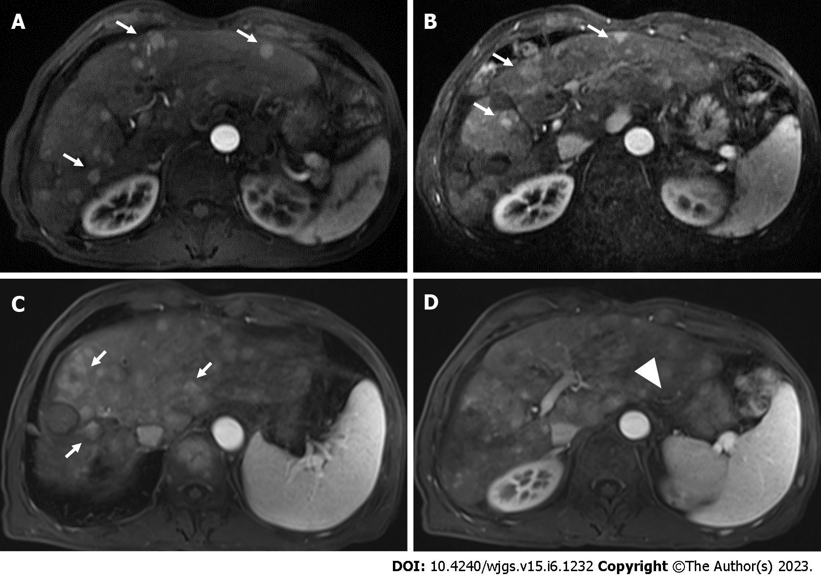
Figure 1 Pseudoaneurysm bleeding after atezolizumab–bevacizumab treatment.
A: Magnetic resonance imaging (MRI) of the abdomen revealed multifocal enhancing hepatocellular carcinoma lesions in the liver (white arrows); B: After transcatheter arterial chemoembolization and lenvatinib treatment, MRI showed disease progression (white arrows); C and D: After three cycles of atezolizumab plus bevacizumab, MRI showed progressive disease (white arrows) but no remarkable gastroesophageal varices (white arrowhead).
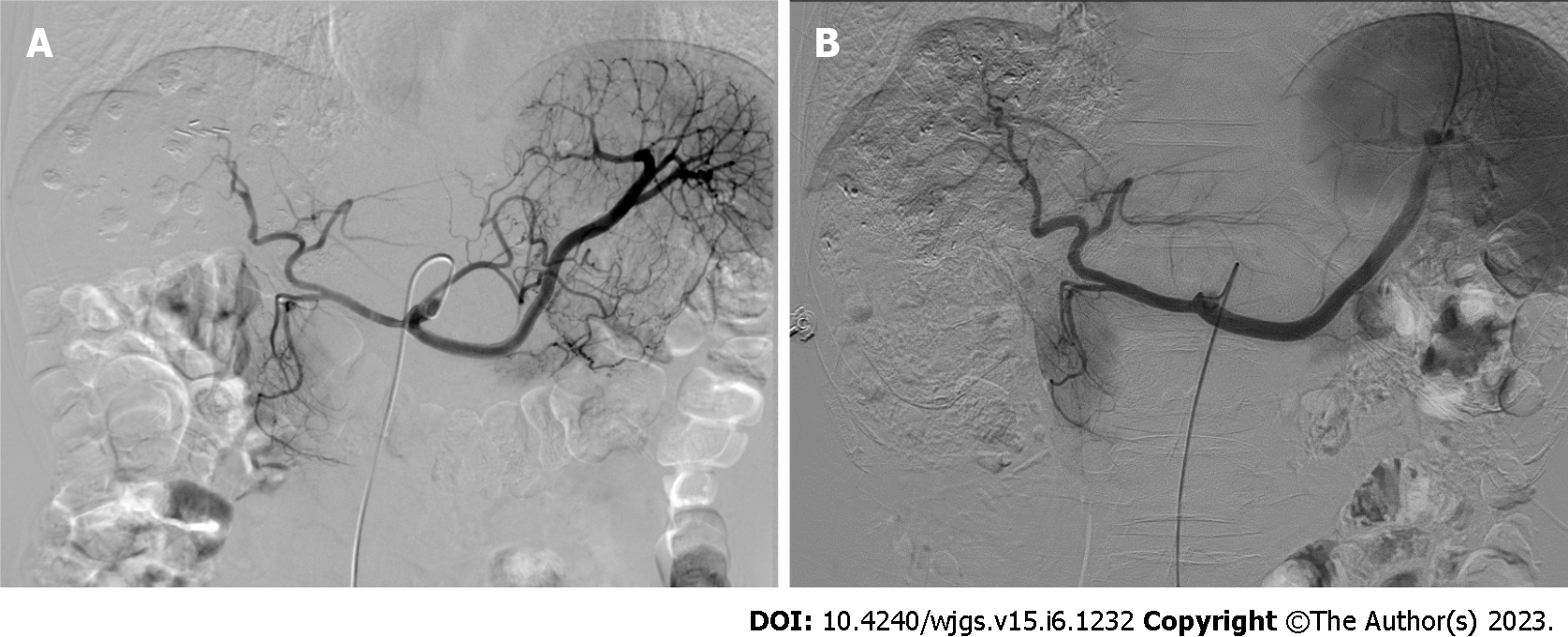
Figure 2 Selective digital subtraction angiography showed no extravasation of contrast medium in the gastric area.
A: Digital subtraction angiography in March 2021; B: Digital subtraction angiography in September 2021.
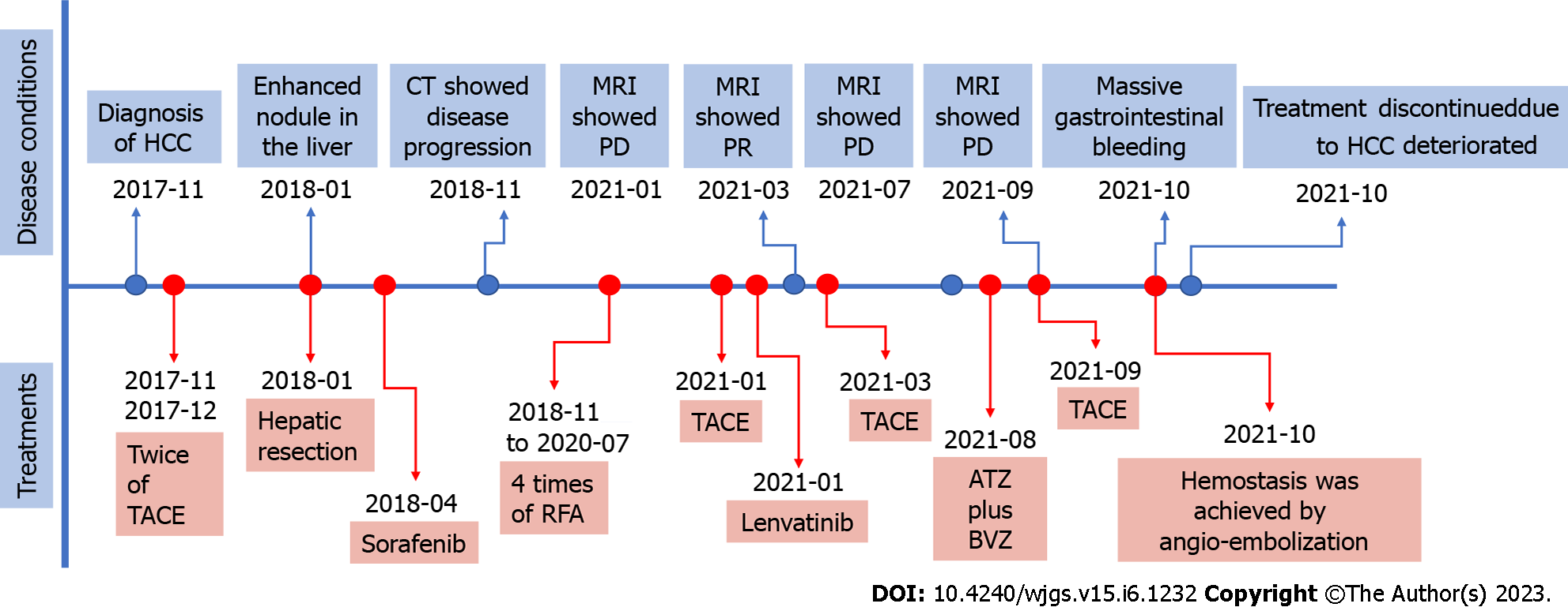
Figure 3 Treatment and disease status timeline.
HCC: Hepatocellular carcinoma; TACE: Transcatheter arterial chemoembolization; CT: Computed tomography; RFA: Radiofrequency ablation; MRI: Magnetic resonance imaging; PD: Progressive disease; PR: Partial response; ATZ: Atezolizumab; BVZ: Bevacizumab.
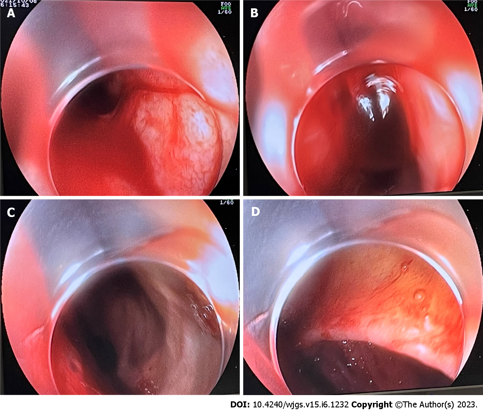
Figure 4 The findings of gastrointestinal endoscopy performed by the time of massive upper gastrointestinal bleeding.
Visualization was limited because of massive fresh blood and blood clots within the stomach. A-C: No remarkable esophageal varices or red wale signs were observed in esophagus and fundus of stomach; D: No recurrence of gastric carcinoma or anastomotic bleeding was detected.
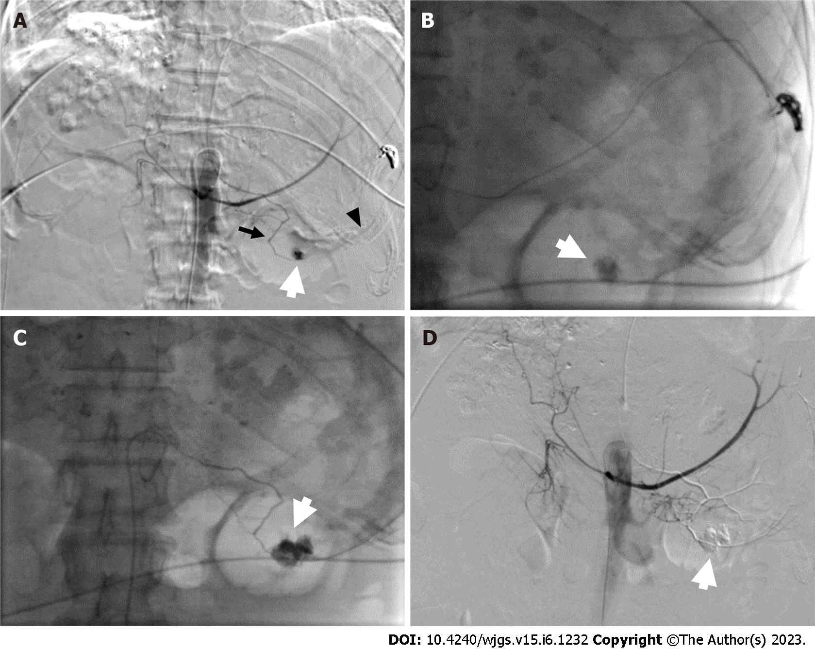
Figure 5 Emergency angiography.
A: Selective digital subtraction angiography of the celiac trunk showed extravasation of contrast medium (white arrow) from the inferior splenic artery (black arrow), a branch of the left gastric artery (black arrow head), and a pseudoaneurysm of the gastric artery; B and C: The pseudoaneurysm (white arrow) was embolized using liquid glue and lipiodol; D: After embolization, the pseudoaneurysm (white arrow) and active bleeding were no longer visible.
- Citation: Pang FW, Chen B, Peng DT, He J, Zhao WC, Chen TT, Xie ZG, Deng HH. Massive bleeding from a gastric artery pseudoaneurysm in hepatocellular carcinoma treated with atezolizumab plus bevacizumab: A case report. World J Gastrointest Surg 2023; 15(6): 1232-1239
- URL: https://www.wjgnet.com/1948-9366/full/v15/i6/1232.htm
- DOI: https://dx.doi.org/10.4240/wjgs.v15.i6.1232


