©The Author(s) 2023.
World J Gastrointest Surg. Jun 27, 2023; 15(6): 1159-1168
Published online Jun 27, 2023. doi: 10.4240/wjgs.v15.i6.1159
Published online Jun 27, 2023. doi: 10.4240/wjgs.v15.i6.1159
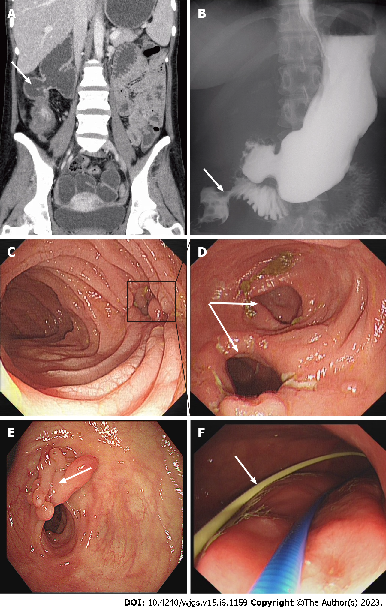
Figure 1 The imaging examinations of patients with an internal fistula between the duodenum and colon.
A: The abdominal computed tomography image showed the existence of an internal fistula between the duodenum and colon; B: The gastrointestinal radiography showed the existence of an internal fistula between the duodenum and colon; C: The ileocecus can be accessed through the internal duodenal fistula under gastroscopy; D: The internal fistula between the duodenum and colon under colonoscopy; E: The placement of jejunal nutrition tube under gastroscope.
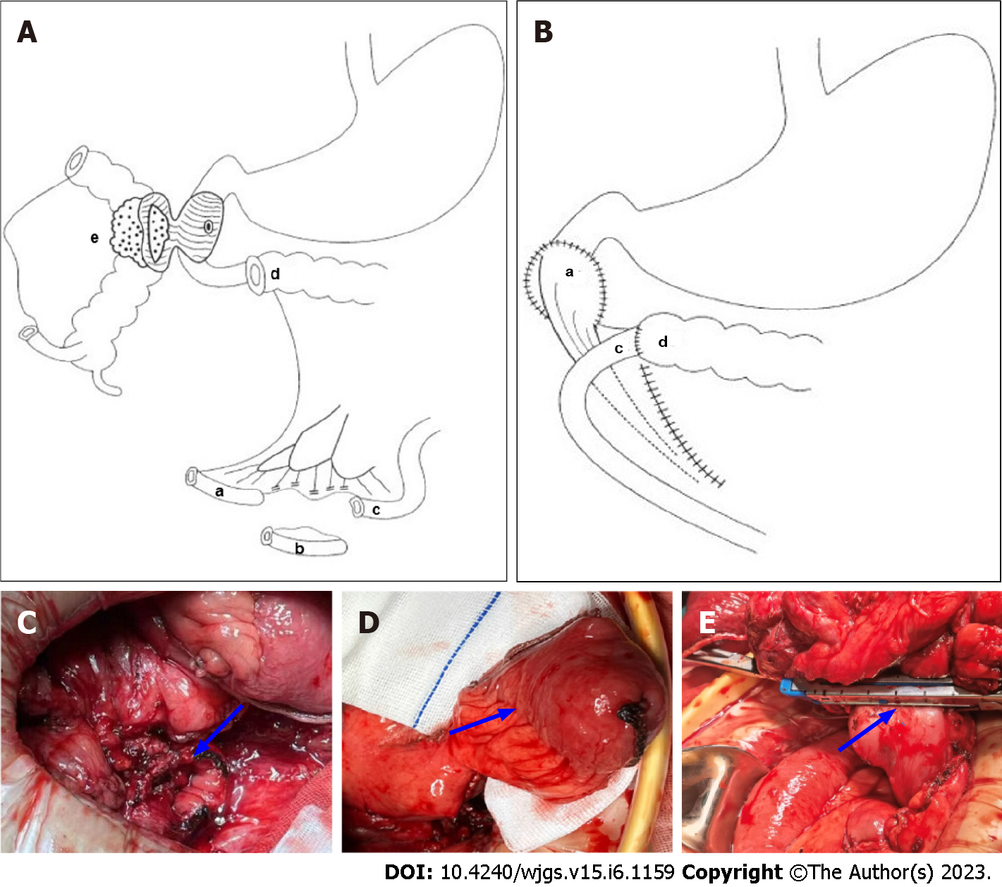
Figure 2 Schematic diagram of pedicled terminal ileum flap closure of the duodenal defect.
A: Pedicled terminal ileum (a), ileum (b), proximal ileum (c), transverse colon (d), Crohn's disease lesions (e); B: Duodenal defect repaired with a pedicled terminal ileal flap (a), terminal ileum (c), transverse colon (d); C: The duodenal defect was larger than 3 cm in diameter; D: Pedicled terminal ileum; E: Direct closure by mechanical stapling of duodenal defects was performed when the duodenal defect was ≤ 3 cm in diameter.
- Citation: Yang LC, Wu GT, Wu Q, Peng LX, Zhang YW, Yao BJ, Liu GL, Yuan LW. Surgical management of duodenal Crohn's disease. World J Gastrointest Surg 2023; 15(6): 1159-1168
- URL: https://www.wjgnet.com/1948-9366/full/v15/i6/1159.htm
- DOI: https://dx.doi.org/10.4240/wjgs.v15.i6.1159


