©The Author(s) 2023.
World J Gastrointest Surg. Mar 27, 2023; 15(3): 440-449
Published online Mar 27, 2023. doi: 10.4240/wjgs.v15.i3.440
Published online Mar 27, 2023. doi: 10.4240/wjgs.v15.i3.440
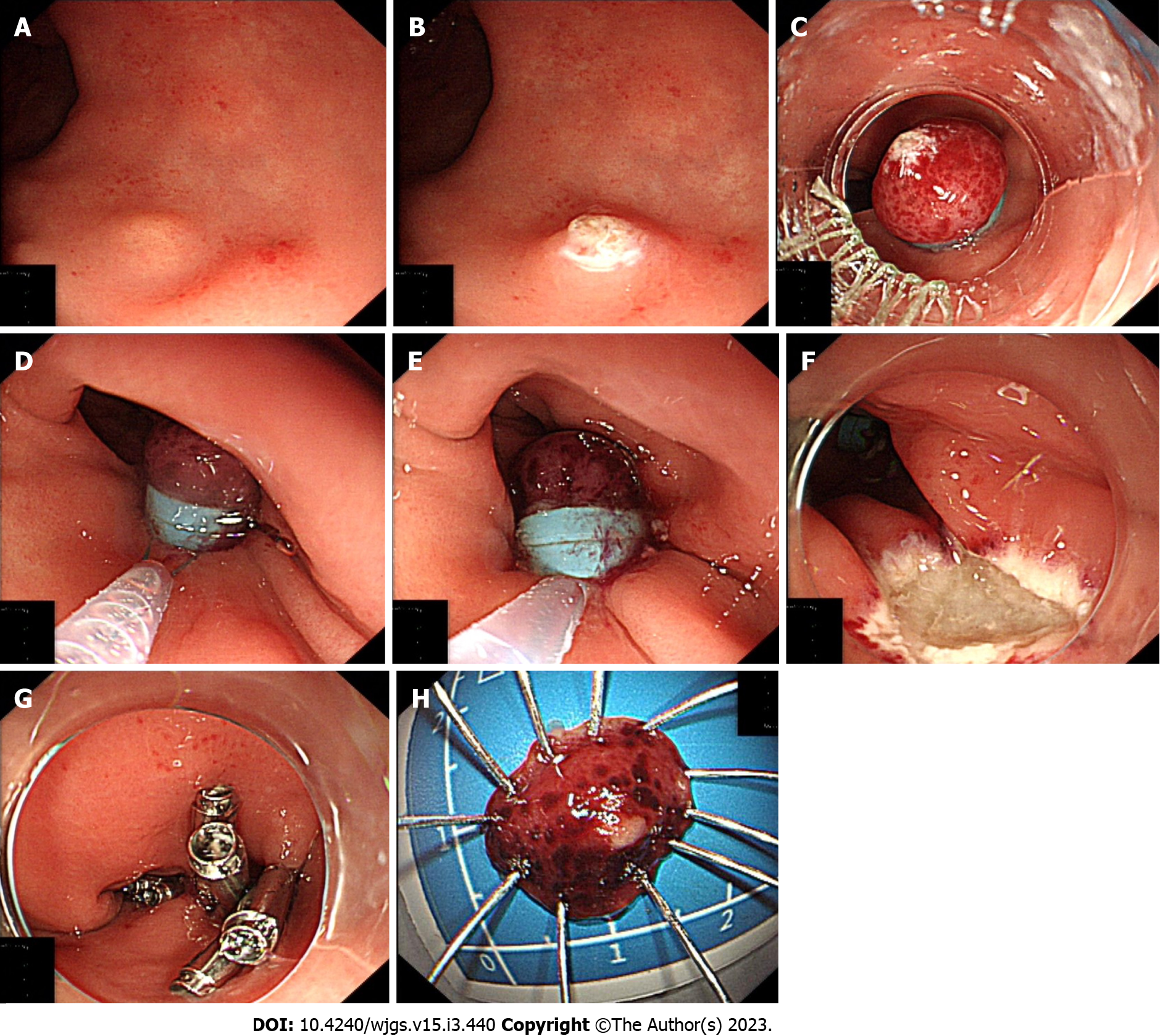
Figure 1 Utilizing Endoscopic mucosal resection with double band ligation to remove the rectal neuroendocrine tumors.
A: Endoscopy showing a rectal neuroendocrine tumors; B: Marking dots were made approximately 2-3 mm on the lesion with an electric snare tip; C: When the lesion was sucked into the ligating device, the first band was deployed to ligate the lesion; D: The second band was deployed below the first one after endoscopic suctioning of the tumor into the cap; E: The resection of the lesion was performed via electrocautery below the second band; F: Wound after resection; G: The wound was closed with clips; H: Endoscopic resection of the intact tumor and the fully flattened specimen.
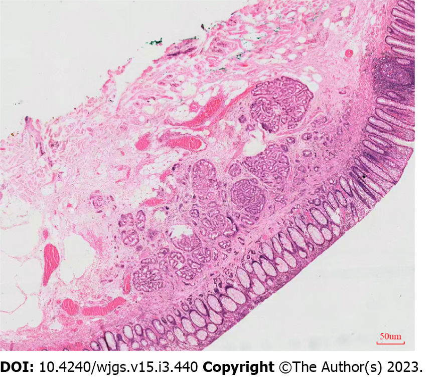
Figure 2 Pathological examination reveals a grade 1 neuroendocrine tumors with negative vertical and lateral margins.
- Citation: Huang JL, Gan RY, Chen ZH, Gao RY, Li DF, Wang LS, Yao J. Endoscopic mucosal resection with double band ligation versus endoscopic submucosal dissection for small rectal neuroendocrine tumors. World J Gastrointest Surg 2023; 15(3): 440-449
- URL: https://www.wjgnet.com/1948-9366/full/v15/i3/440.htm
- DOI: https://dx.doi.org/10.4240/wjgs.v15.i3.440


