©The Author(s) 2022.
World J Gastrointest Surg. Aug 27, 2022; 14(8): 855-861
Published online Aug 27, 2022. doi: 10.4240/wjgs.v14.i8.855
Published online Aug 27, 2022. doi: 10.4240/wjgs.v14.i8.855
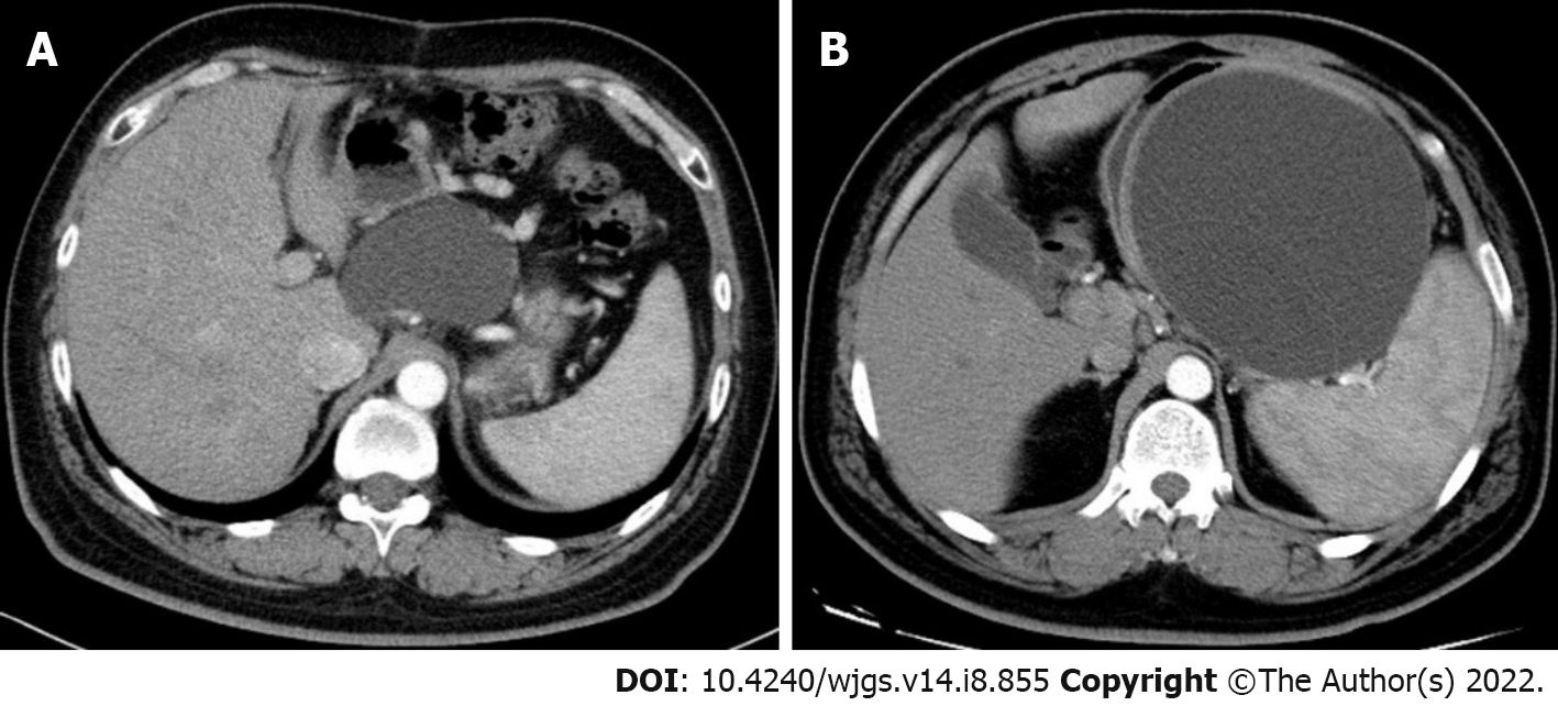
Figure 1 Preoperative images of contrast-enhanced computed tomography.
A: Preoperative contrast-enhanced computed tomography (CECT) image of the first patient showed a cystic lesion in the body of the pancreas, with a size of 7.93 cm × 6.13 cm; B: Preoperative CECT image of the second patient showed a cystic lesion with a maximum diameter of 14 cm.
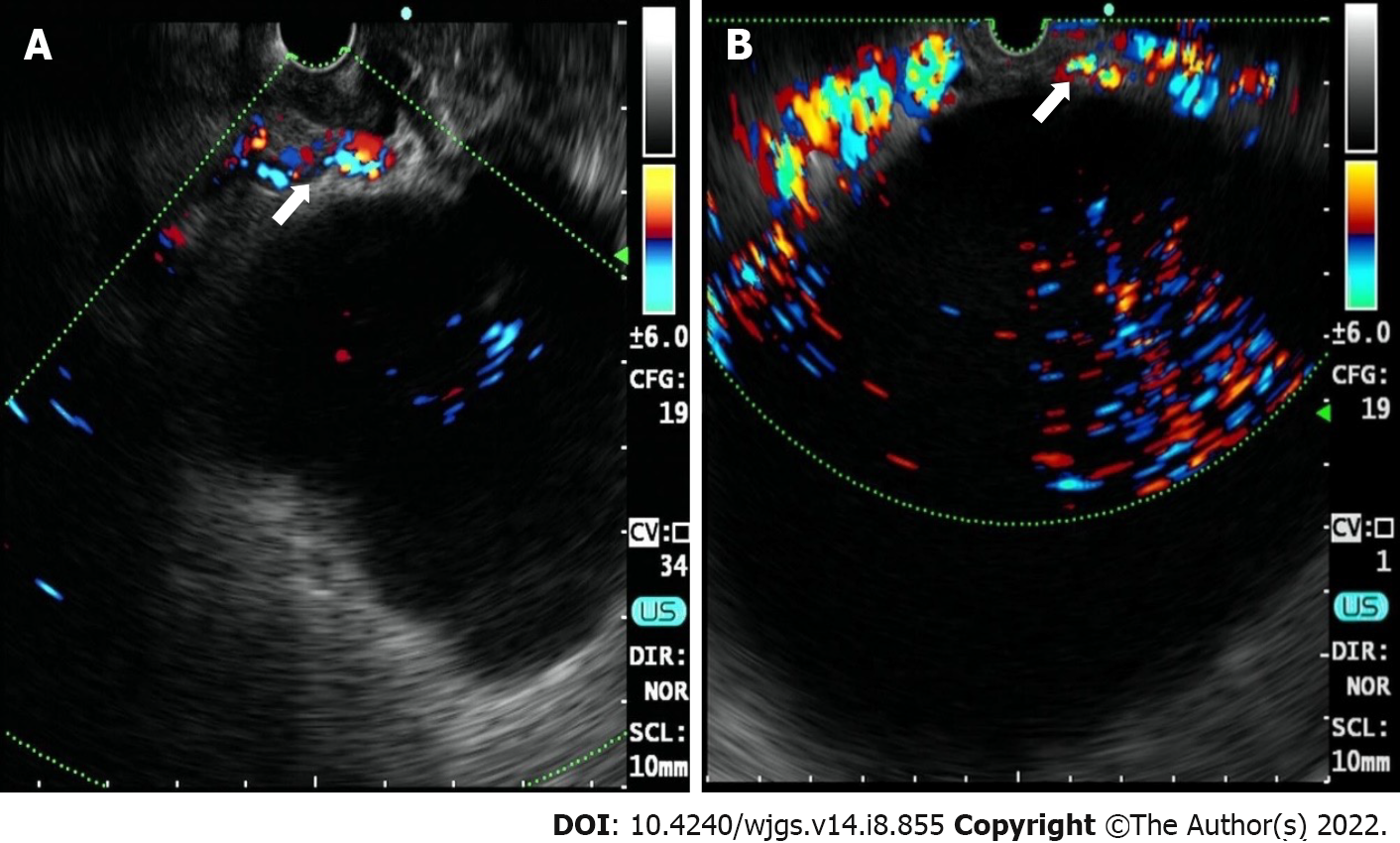
Figure 2 Multiple vasculature (white arrow) detected by Doppler endoscopic ultrasound.
A: Endoscopic ultrasound (EUS) imaging of the first patient; B: EUS imaging of the second patient.
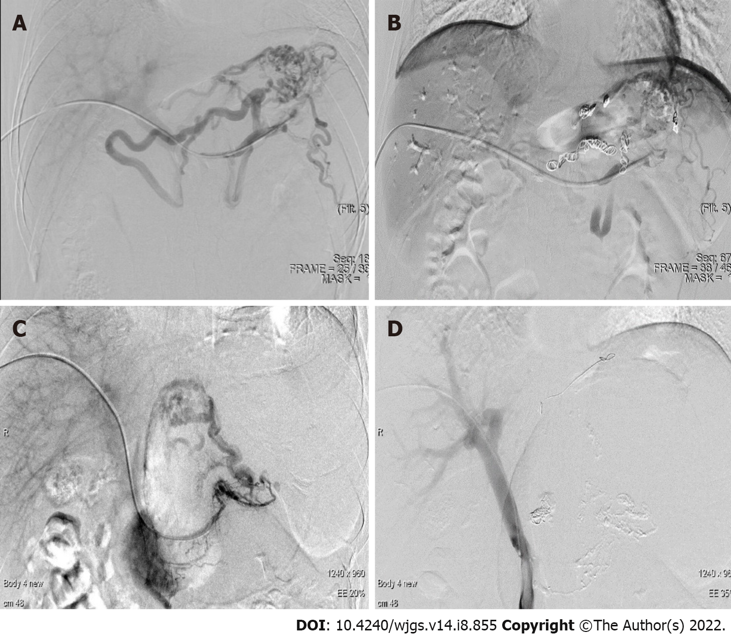
Figure 3 Typical imaging of interventional radiology.
A: Angiogram of the first patient prior to coil embolization; B: Angiogram of the first patient after coil embolization; C: Angiogram of the second patient prior to coil embolization; D: Angiogram of the second patient after coil embolization.
- Citation: Xu N, Li LS, Yue WY, Zhao DQ, Xiang JY, Zhang B, Wang PJ, Cheng YX, Linghu EQ, Chai NL. Interventional radiology followed by endoscopic drainage for pancreatic fluid collections associated with high bleeding risk: Two case reports. World J Gastrointest Surg 2022; 14(8): 855-861
- URL: https://www.wjgnet.com/1948-9366/full/v14/i8/855.htm
- DOI: https://dx.doi.org/10.4240/wjgs.v14.i8.855


