Copyright
©The Author(s) 2022.
World J Gastrointest Surg. Jun 27, 2022; 14(6): 611-620
Published online Jun 27, 2022. doi: 10.4240/wjgs.v14.i6.611
Published online Jun 27, 2022. doi: 10.4240/wjgs.v14.i6.611
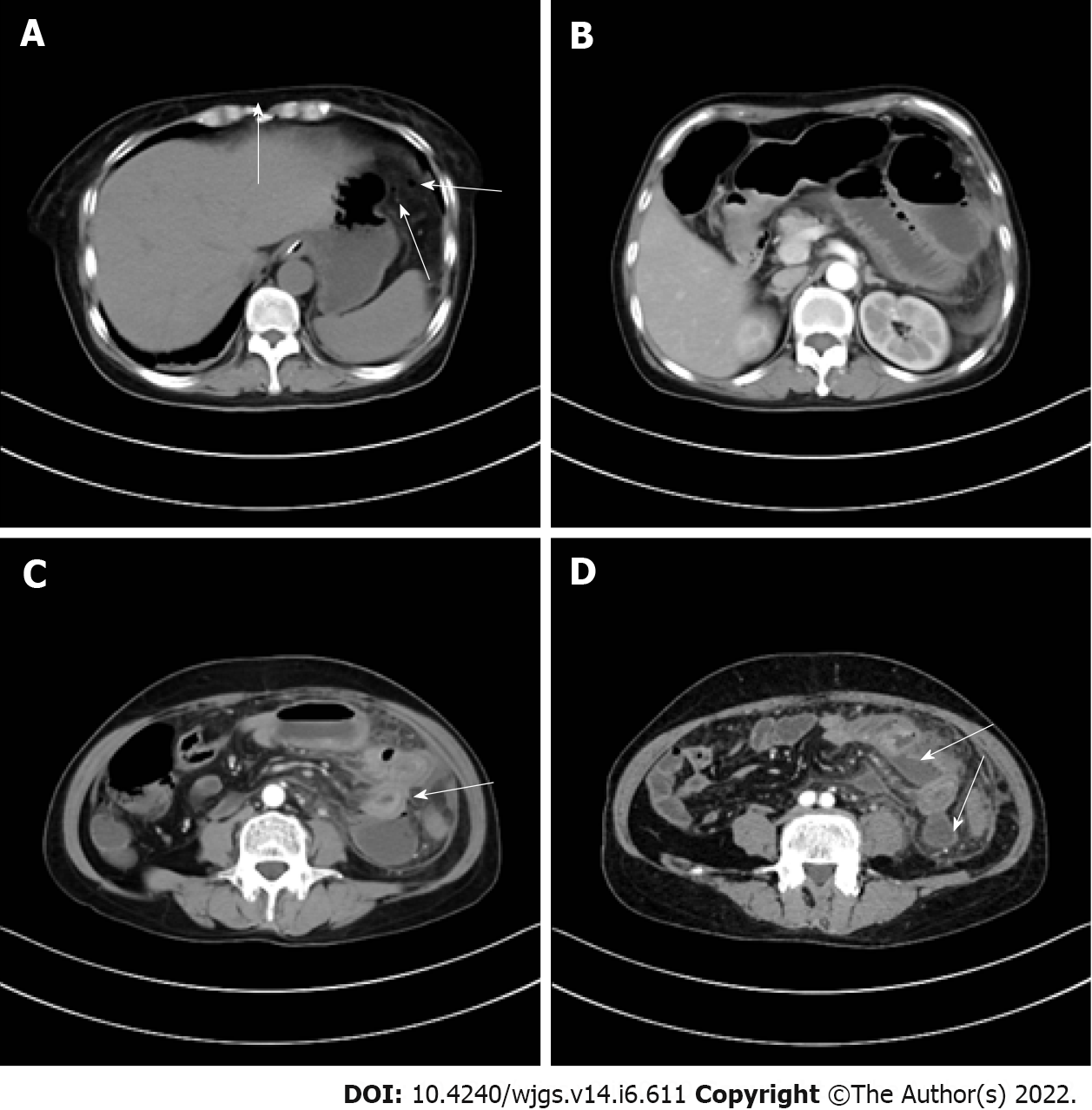
Figure 1 Preoperative computed tomography scan findings.
A: There are small air bubbles scattered under the left diaphragm (indicated by white arrow); B: The small intestinal lumen in the upper left abdomen is dilated with gas and fluid accumulation, showing multiple fluid-gas level changes; C: The intestinal wall presents edematous thickening (indicated by white arrow), and the density of local mesentery increases; D: Multiple abscesses can be seen between the intestinal lumen (indicated by white arrow).
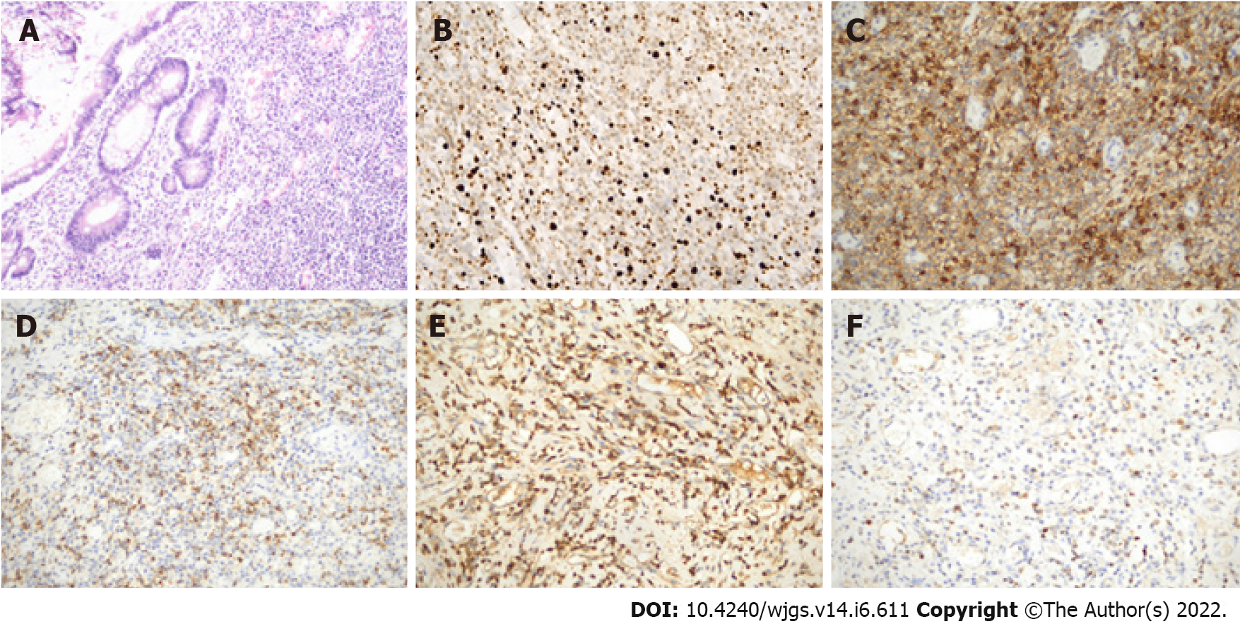
Figure 2 Histopathological examination of extramedullary plasmacytoma of small intestine.
Microscopic view of the resected extramedullary plasmacytoma originating from small intestine. A: Hematoxylin and eosin staining, magnification × 100; B: Ki67, magnification × 200; C: CD38, magnification × 200; D: CD138, magnification × 200; E: Kappa, magnification × 200; F: Lambda, magnification × 200.
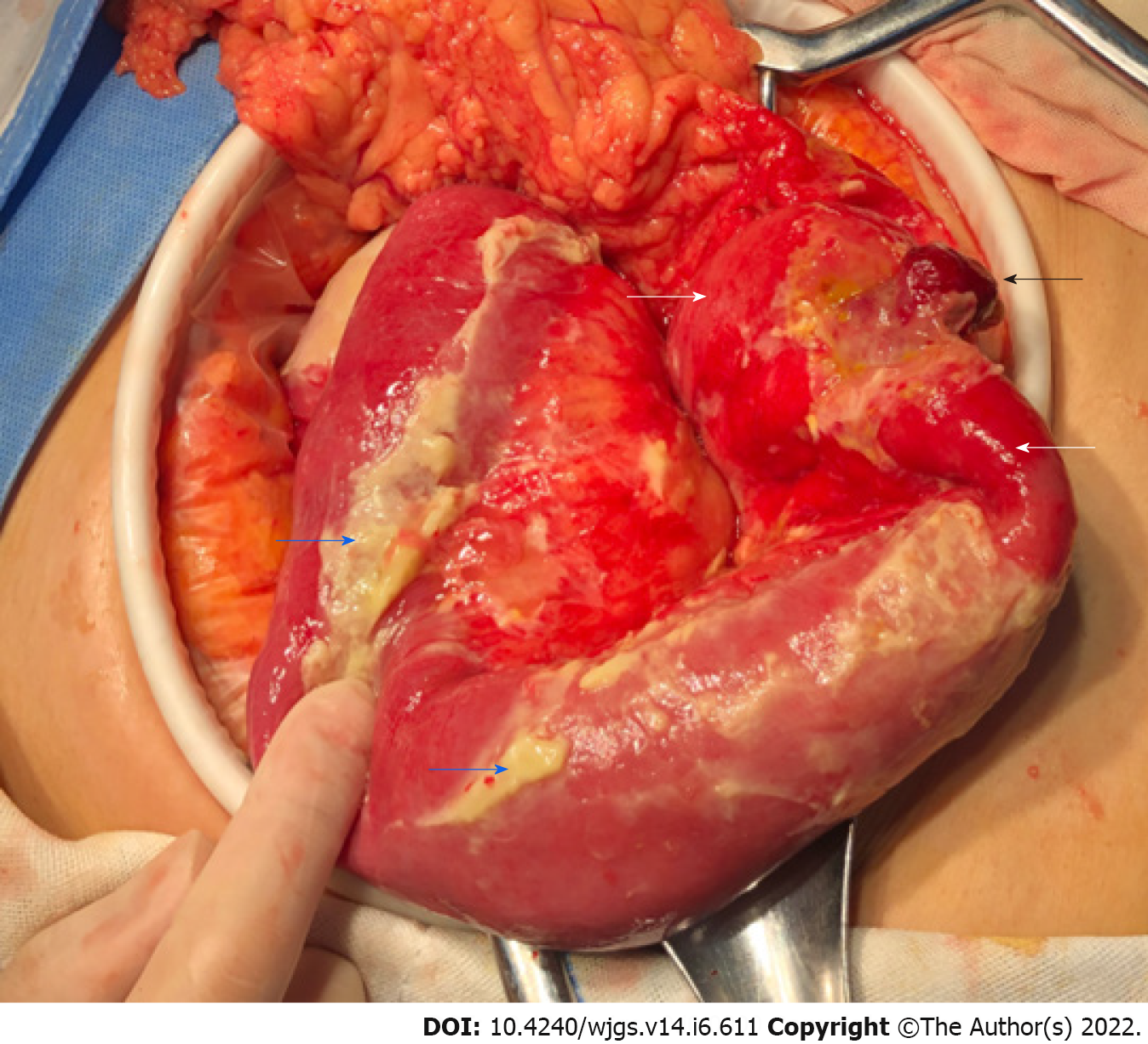
Figure 3 Intra-operative findings.
The small intestinal serosa has a dark red polyp-like protrusion (black arrow) with a perforation about 0.5 cm in diameter at the top. The local intestinal wall presents hyperemia, edema and thickening (white arrow). The surface of the surrounding small intestine is covered with a large amount of purulent material (blue arrow).
- Citation: Wang KW, Xiao N. Intestinal perforation with abdominal abscess caused by extramedullary plasmacytoma of small intestine: A case report and literature review. World J Gastrointest Surg 2022; 14(6): 611-620
- URL: https://www.wjgnet.com/1948-9366/full/v14/i6/611.htm
- DOI: https://dx.doi.org/10.4240/wjgs.v14.i6.611


