©The Author(s) 2022.
World J Gastrointest Surg. Apr 27, 2022; 14(4): 352-361
Published online Apr 27, 2022. doi: 10.4240/wjgs.v14.i4.352
Published online Apr 27, 2022. doi: 10.4240/wjgs.v14.i4.352
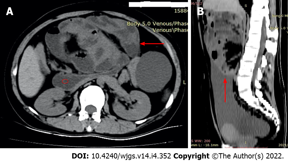
Figure 1 Computerized tomography of transverse plane and sagittal plane.
A: Computerized tomography (CT) of transverse plane: A conglomerate of multiple intestinal loops encapsulated in a thick sac-like membrane (arrow), and dilated duodenum (red cycle); B: CT of sagittal plane: Epigastric mass floating in ascites.
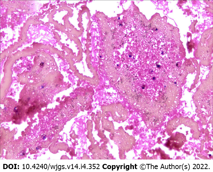
Figure 2 Exfoliative cytology of ascites (hematoxylin and eosin stain, × 40).
A large number of red blood cells, including scattered inflammatory cells.
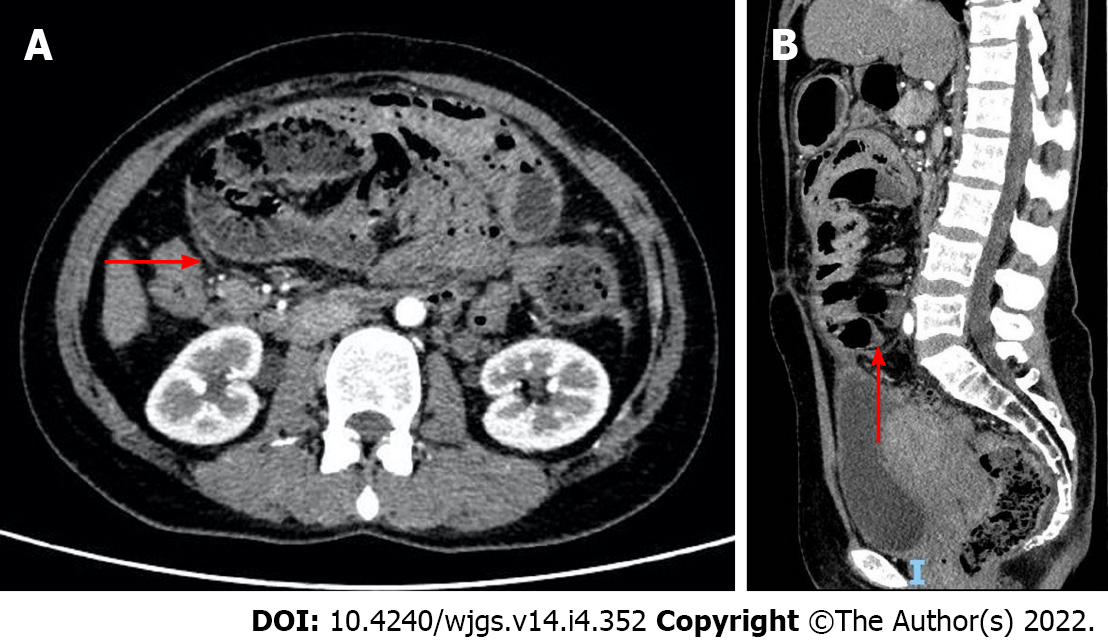
Figure 3 Contrast-enhanced computerized tomography of transverse plane and sagittal plane.
A: Transverse plane; B: Sagittal plane. Intestinal loops were encapsulated in a thick sac-like membrane (arrow).
- Citation: Deng P, Xiong LX, He P, Hu JH, Zou QX, Le SL, Wen SL. Surgical timing for primary encapsulating peritoneal sclerosis: A case report and review of literature. World J Gastrointest Surg 2022; 14(4): 352-361
- URL: https://www.wjgnet.com/1948-9366/full/v14/i4/352.htm
- DOI: https://dx.doi.org/10.4240/wjgs.v14.i4.352


