©The Author(s) 2022.
World J Gastrointest Surg. Mar 27, 2022; 14(3): 260-267
Published online Mar 27, 2022. doi: 10.4240/wjgs.v14.i3.260
Published online Mar 27, 2022. doi: 10.4240/wjgs.v14.i3.260
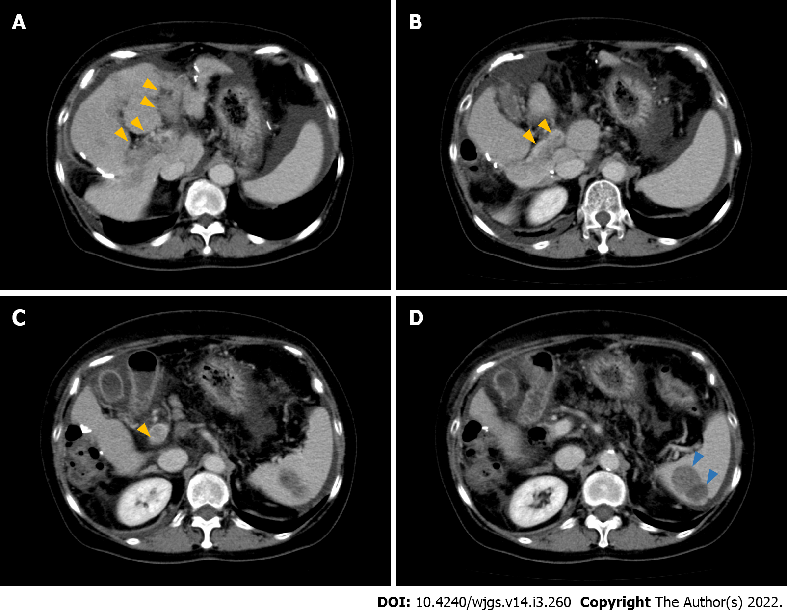
Figure 1 Radiological findings of hepatocellular carcinoma with portal vein tumor thrombosis and splenic metastasis.
A: Hypervascular lesion in the left and right anterior portal branches (yellow arrows) suggesting portal vein tumor thrombosis. Ascites are located around the spleen. Dynamic computed tomography (CT), portal phase; B and C: Hypervascular lesions in the main portal branch (yellow arrows). Dynamic CT, portal phase; D: Heterogenic, largely a hypodense lesion with high contrast enhancement in the lower pole of the spleen (blue arrows). Dynamic CT, portal phase.
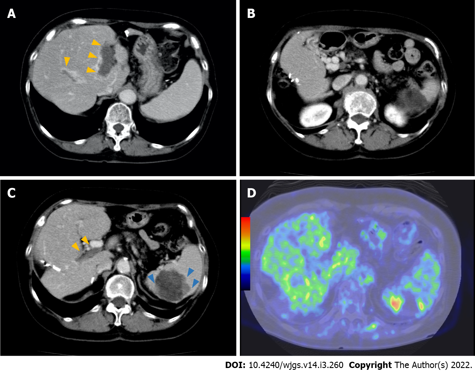
Figure 2 Radiological findings after the lenvatinib treatment.
A: Portal vein tumor thrombus (PVTT) becomes hypovascular (yellow arrows) 10 mo after the administration of lenvatinib. Dynamic computed tomography (CT), portal phase; B: The main portal vein is hypovascular, suggesting the organization of PVTT 10 mo after the administration of lenvatinib. Numerous collateral veins are seen around the portal vein. Dynamic CT, portal phase; C: PVTT remains hypovascular (yellow arrows), whereas hypervascular lesions increase the peripheral lesions of the spleen metastases (blue arrows) 8 mo after the cessation of lenvatinib. Dynamic CT, portal phase; D: Fluorodeoxyglucose (FDG) uptakes in the lower pole of the spleen (blue arrows) corresponding to hypervascular lesions on CT. FDG-positron emission tomography.
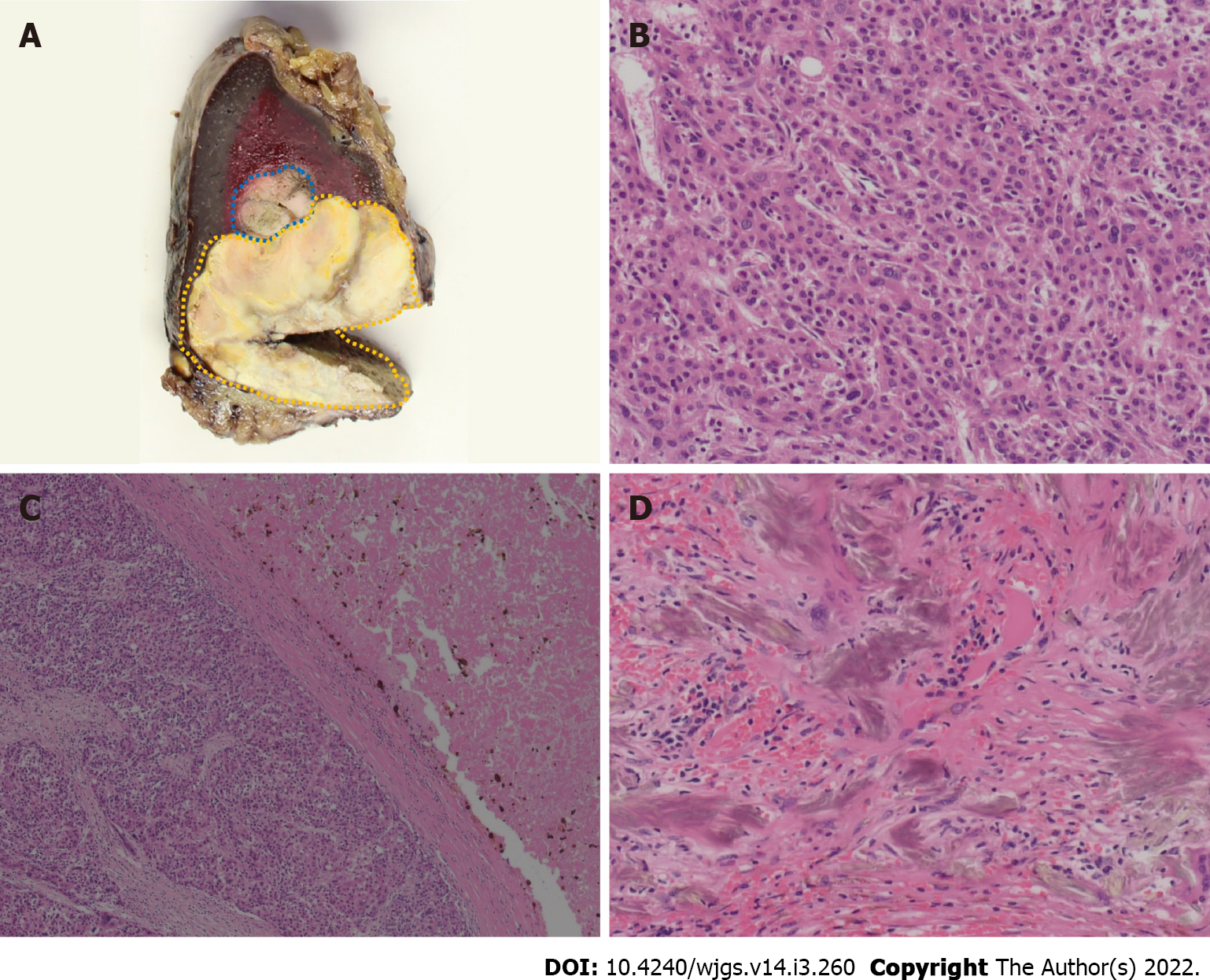
Figure 3 Pathological findings of metastatic splenic lesions.
A: Macroscopic finding shows that splenic lesions surrounded by fibrous capsule, and a border part (blue area) is distinguished from other parts (yellow area) with its color, suggesting viable lesions; B: Microscopic finding of viable tumor lesion shows moderately to poorly differentiated hepatocellular carcinoma. Hematoxylin-eosin stain, high-power field (× 200); C: Microscopic findings of mixed component of viable cells and necrotic tissue demonstrated that coagulative and partially liquefactive necrosis (right-side) is surrounded by fibrous capsule, and viable cells (left-side). Hematoxylin-eosin stain, low-power field (× 50); D: Gamma-Gandy bodies shown in the splenic lesions, suggesting previous history of portal hypertension due to portal vein tumor thrombosis. Hematoxylin-eosin stain, high-power field (× 200).
- Citation: Endo Y, Shimazu M, Sakuragawa T, Uchi Y, Edanami M, Sunamura K, Ozawa S, Chiba N, Kawachi S. Successful treatment with laparoscopic surgery and sequential multikinase inhibitor therapy for hepatocellular carcinoma: A case report. World J Gastrointest Surg 2022; 14(3): 260-267
- URL: https://www.wjgnet.com/1948-9366/full/v14/i3/260.htm
- DOI: https://dx.doi.org/10.4240/wjgs.v14.i3.260


