©The Author(s) 2022.
World J Gastrointest Surg. Dec 27, 2022; 14(12): 1418-1424
Published online Dec 27, 2022. doi: 10.4240/wjgs.v14.i12.1418
Published online Dec 27, 2022. doi: 10.4240/wjgs.v14.i12.1418
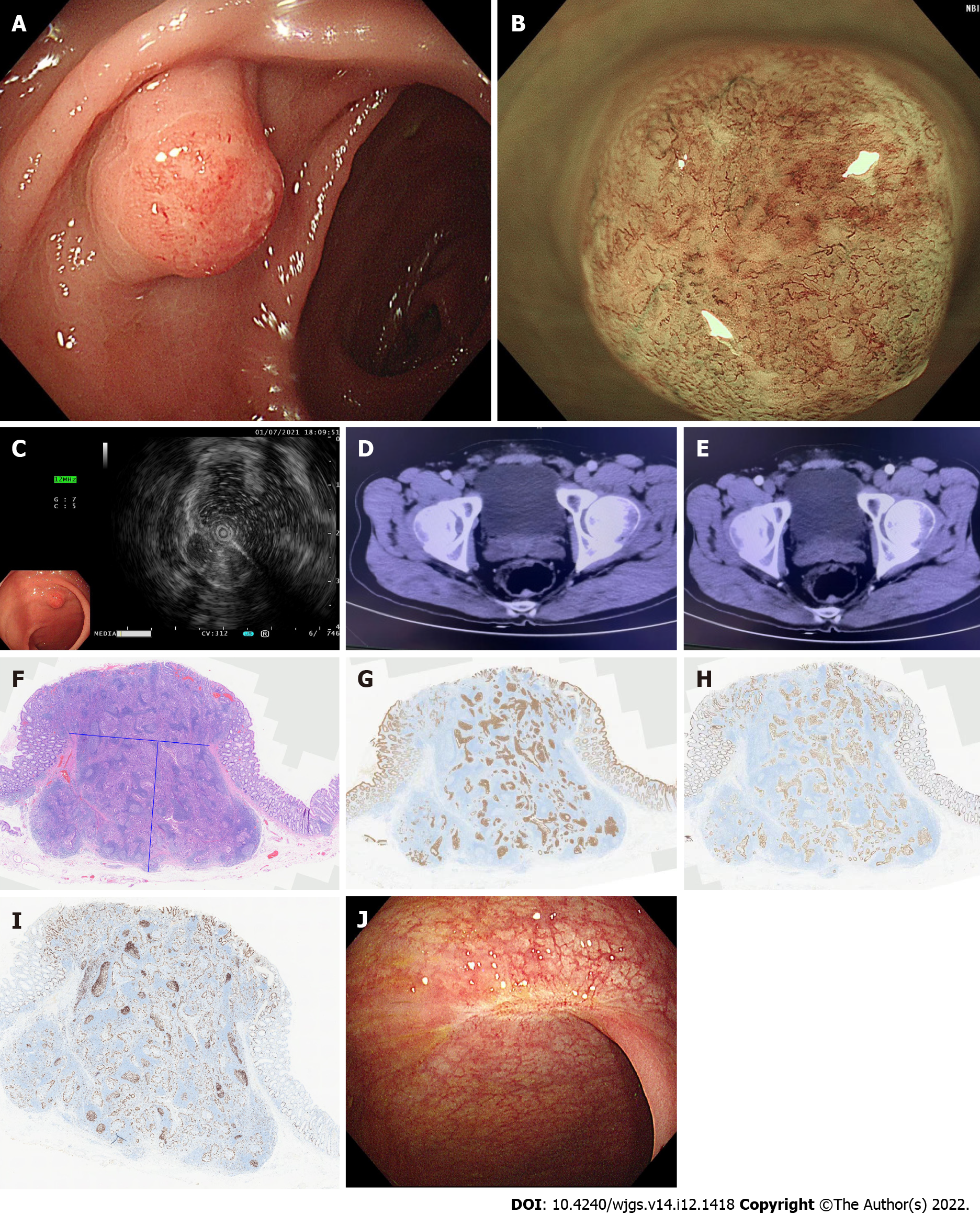
Figure 1 Clinical data of the patient with rectal tubular adenoma and submucosal pseudoinvasion.
A: Normal colonoscopy showing Is-type lesion; B: Narrow band imaging magnification colonoscopy; C: Ultrasound colonoscopy image; D: Posterior rectal wall in the venous phase showing the lesion; E: Posterior rectal wall in the arterial phase showing the lesion; F: The epithelial tumors seen microscopically are glandular ductal and sieve shaped with a high-grade heterogeneous morphology. The tumors are located in the lymphatic interstitium rich in lymphoid follicles. The lymphoid interstitium is located in the deep mucosal and submucosal layers with smooth and well-defined borders, H&E × 200 magnification; G: Immunohistochemistry CK20 (+), magnification × 200; H: Immunohistochemistry CDX2 (+), magnification × 200; I: Immunohistochemistry Ki67 (30%+), magnification × 200; J: Colonoscopy results after the operation.
- Citation: Chen D, Zhong DF, Zhang HY, Nie Y, Liu D. Rectal tubular adenoma with submucosal pseudoinvasion misdiagnosed as adenocarcinoma: A case report. World J Gastrointest Surg 2022; 14(12): 1418-1424
- URL: https://www.wjgnet.com/1948-9366/full/v14/i12/1418.htm
- DOI: https://dx.doi.org/10.4240/wjgs.v14.i12.1418


