©The Author(s) 2020.
World J Gastrointest Surg. Sep 27, 2020; 12(9): 397-406
Published online Sep 27, 2020. doi: 10.4240/wjgs.v12.i9.397
Published online Sep 27, 2020. doi: 10.4240/wjgs.v12.i9.397
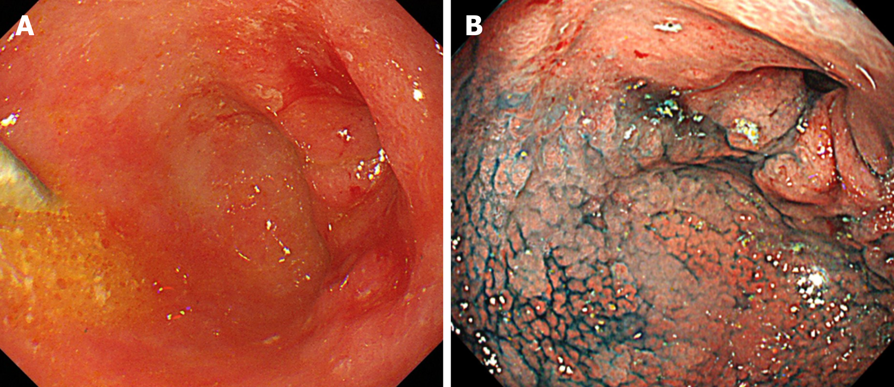
Figure 1 Endoscopic findings.
A: Esophagogastroduodenoscopy revealed a 0-IIc primary tumor with ulcerations surrounding the pyloric ring; B: The lesion was unstained by indigo carmine.
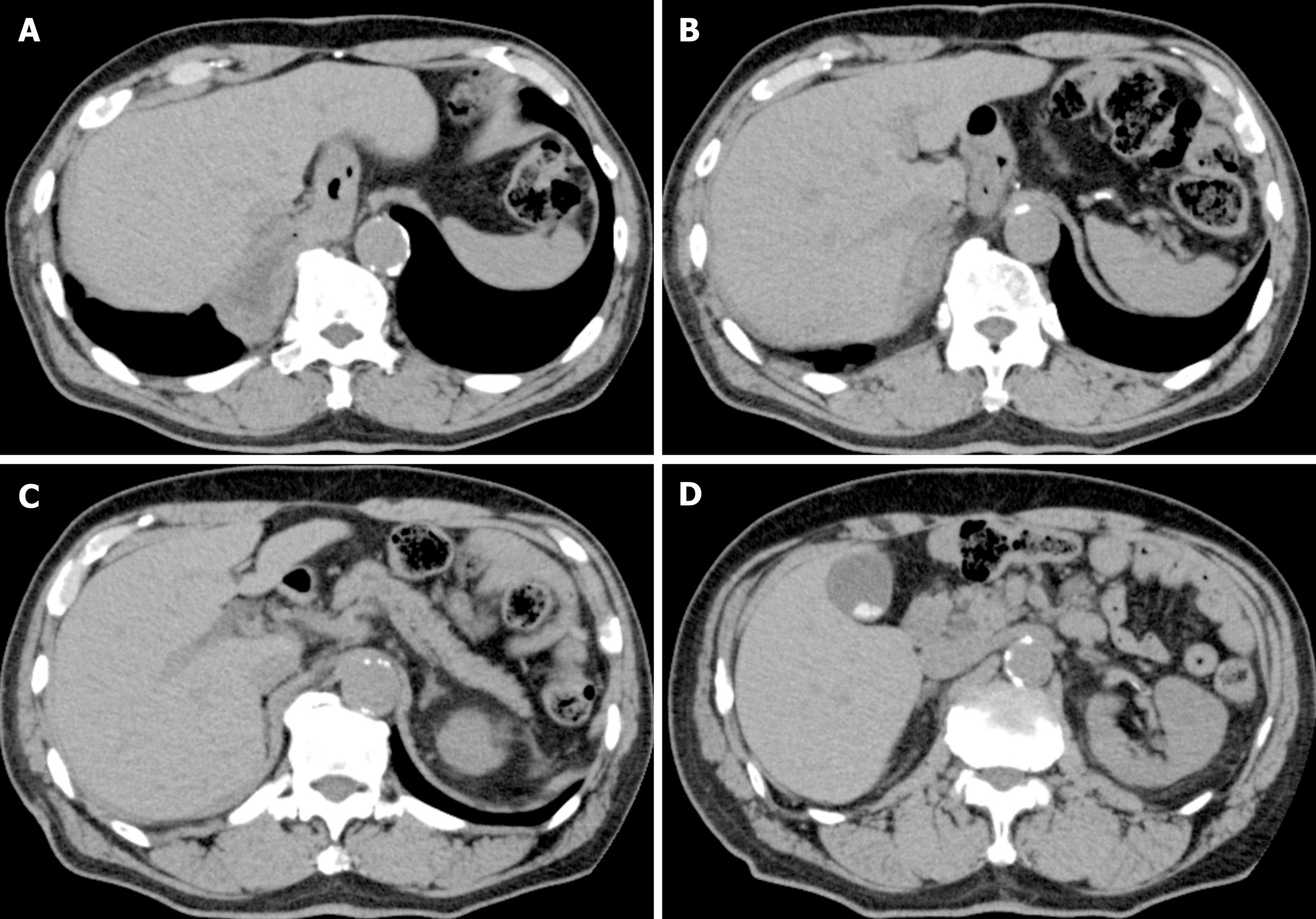
Figure 2 Computed tomography findings.
A-D: Computed tomography was unable to detect the primary tumor, and no obvious nodal metastasis was observed.
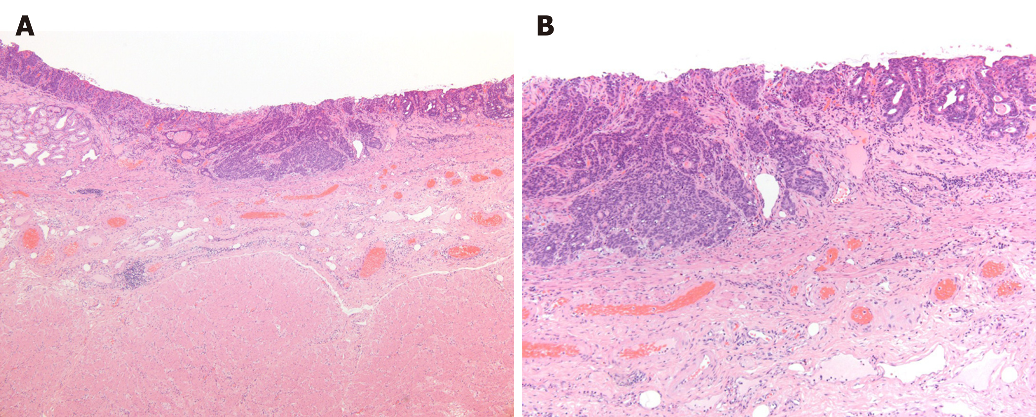
Figure 3 Pathological findings.
Pathological examination of the resected material revealed a moderately to poorly differentiated adenocarcinoma with submucosal invasion and ulcerative scars, but without lymphovascular invasion. A: Hematoxylin and eosin staining results, × 4; B: Hematoxylin and eosin staining results, × 10.
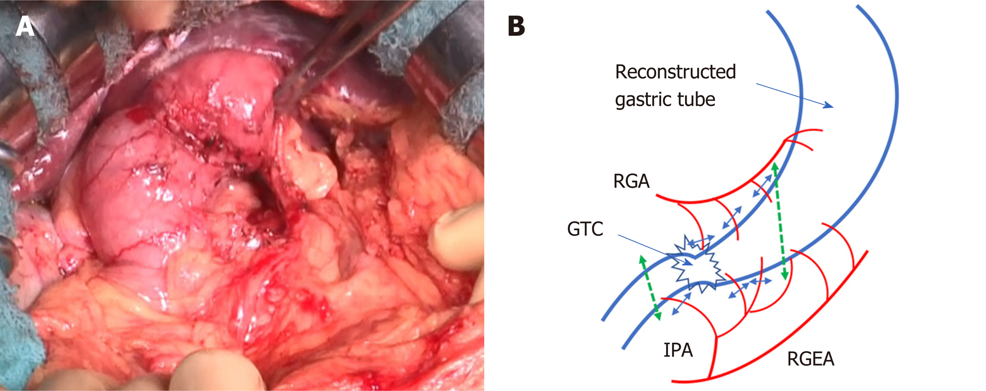
Figure 4 Tumor location.
The peripheral branches to the stomach wall were cut according to the area to be resected, to preserve the main trunks of the right gastric artery and right gastroepiploic artery. Photograph taken during surgery showing the resected branches of the infrapyloric artery. Schematic of the dissection line. GTC: Gastric tube cancer; IPA: Infrapyloric artery; RGA: Right gastric artery; RGEA: Right gastroepiploic artery.
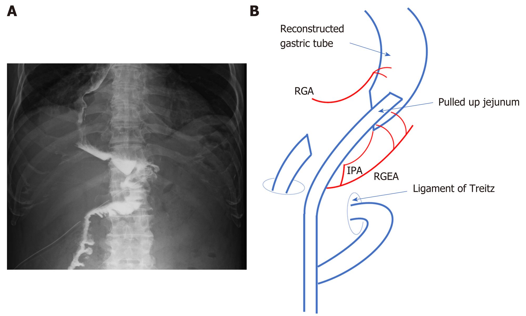
Figure 5 Roux-en-Y reconstruction.
Barium swallow test with antecolic Roux-en-Y reconstruction. Schematic of gastrojejunostomy with antecolic Roux-en-Y reconstruction. IPA: Infrapyloric artery; RGA: Right gastric artery; RGEA: Right gastroepiploic artery.
- Citation: Yura M, Koyanagi K, Adachi K, Hara A, Hayashi K, Tajima Y, Kaneko Y, Fujisaki H, Hirata A, Takano K, Hongo K, Yo K, Yoneyama K, Dehari R, Nakagawa M. Distal gastric tube resection with vascular preservation for gastric tube cancer: A case report and review of literature. World J Gastrointest Surg 2020; 12(9): 397-406
- URL: https://www.wjgnet.com/1948-9366/full/v12/i9/397.htm
- DOI: https://dx.doi.org/10.4240/wjgs.v12.i9.397


