©The Author(s) 2020.
World J Gastrointest Surg. Mar 27, 2020; 12(3): 93-103
Published online Mar 27, 2020. doi: 10.4240/wjgs.v12.i3.93
Published online Mar 27, 2020. doi: 10.4240/wjgs.v12.i3.93
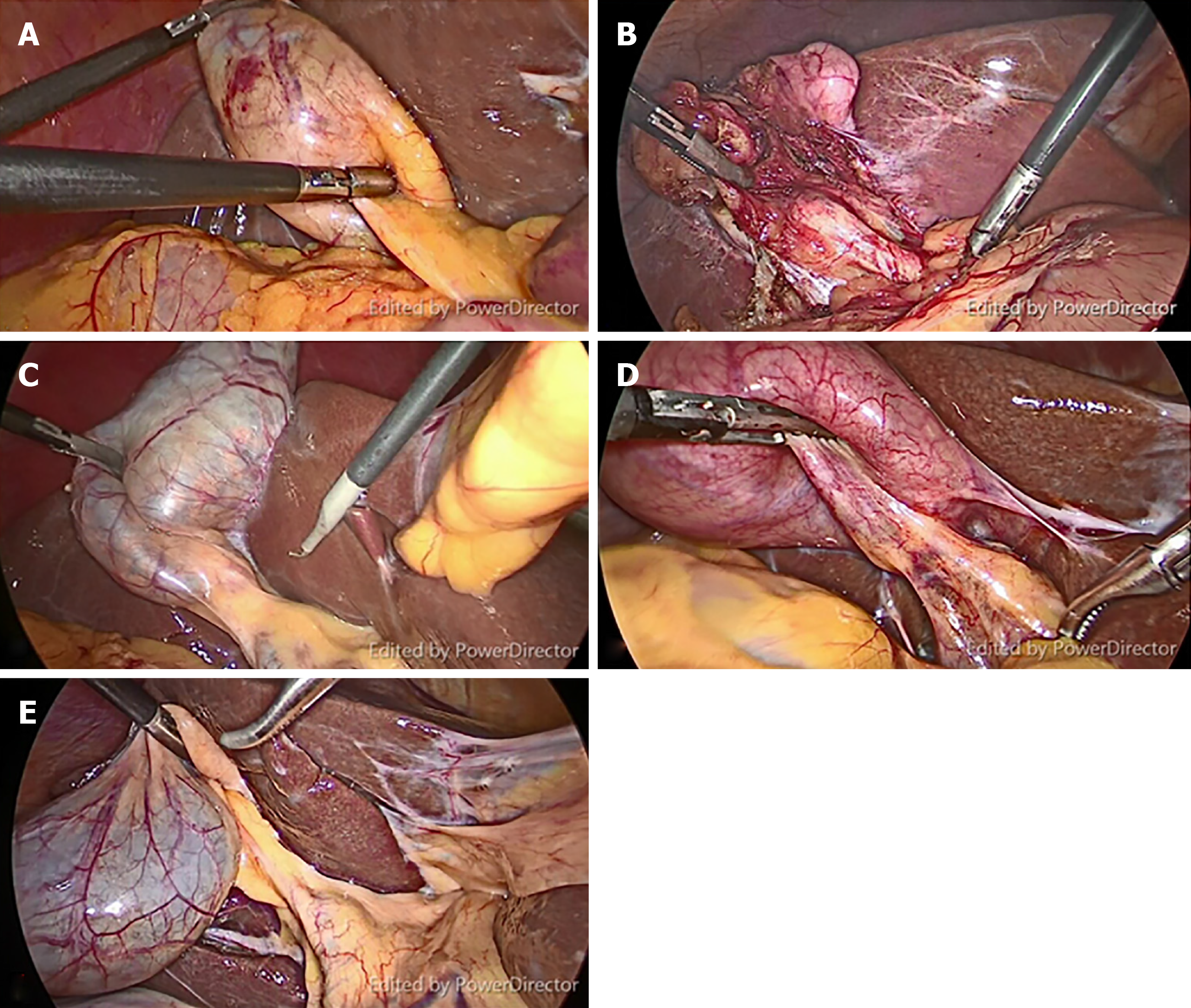
Figure 1 Representative still images from video clips of five patients with different gallbladder diseases.
A: The obese patient with gallbladder polyp; B: Biliary pancreatitis; C: Symptomatic gallstone; D: Acute cholecystitis; E: Gallbladder polyp in a non-obese patient.
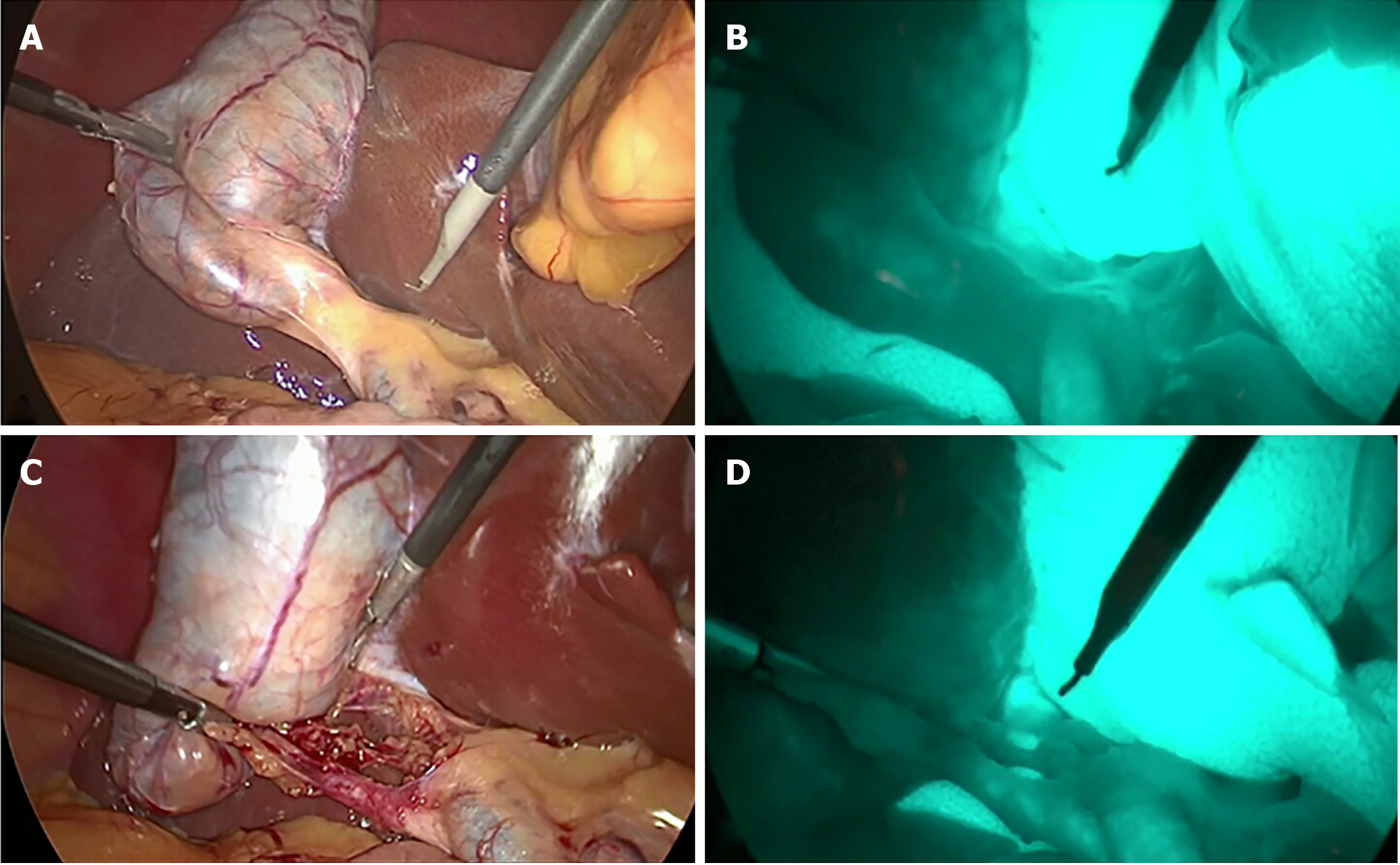
Figure 2 Representative still images from video clips of each phase of procedure.
A: Before dissection of Calot’s triangle without fluorescence cholangiography (FC); B: Before dissection of Calot’s triangle with FC; C: After dissection of Calot’s triangle without FC; D: After dissection of Calot’s triangle with FC.
Figure 3 Study protocol flow chart.
Figure 4 Representative still images from paired structures identified in both with-and without-fluorescence cholangiography phases from before dissection in the same patient.
A: Before dissection of Calot’s triangle without fluorescence cholangiography; B: Before dissection of Calot’s triangle with fluorescence cholangiography. CD: Cystic duct; CBD: Common bile duct.
- Citation: Rungsakulkij N, Thewmorakot S, Suragul W, Vassanasiri W, Tangtawee P, Muangkaew P, Mingphruedhi S, Aeesoa S. Fluorescence cholangiography enhances surgical residents’ biliary delineation skill for laparoscopic cholecystectomies. World J Gastrointest Surg 2020; 12(3): 93-103
- URL: https://www.wjgnet.com/1948-9366/full/v12/i3/93.htm
- DOI: https://dx.doi.org/10.4240/wjgs.v12.i3.93


