Copyright
©The Author(s) 2019.
World J Gastrointest Surg. Apr 27, 2019; 11(4): 237-246
Published online Apr 27, 2019. doi: 10.4240/wjgs.v11.i4.237
Published online Apr 27, 2019. doi: 10.4240/wjgs.v11.i4.237
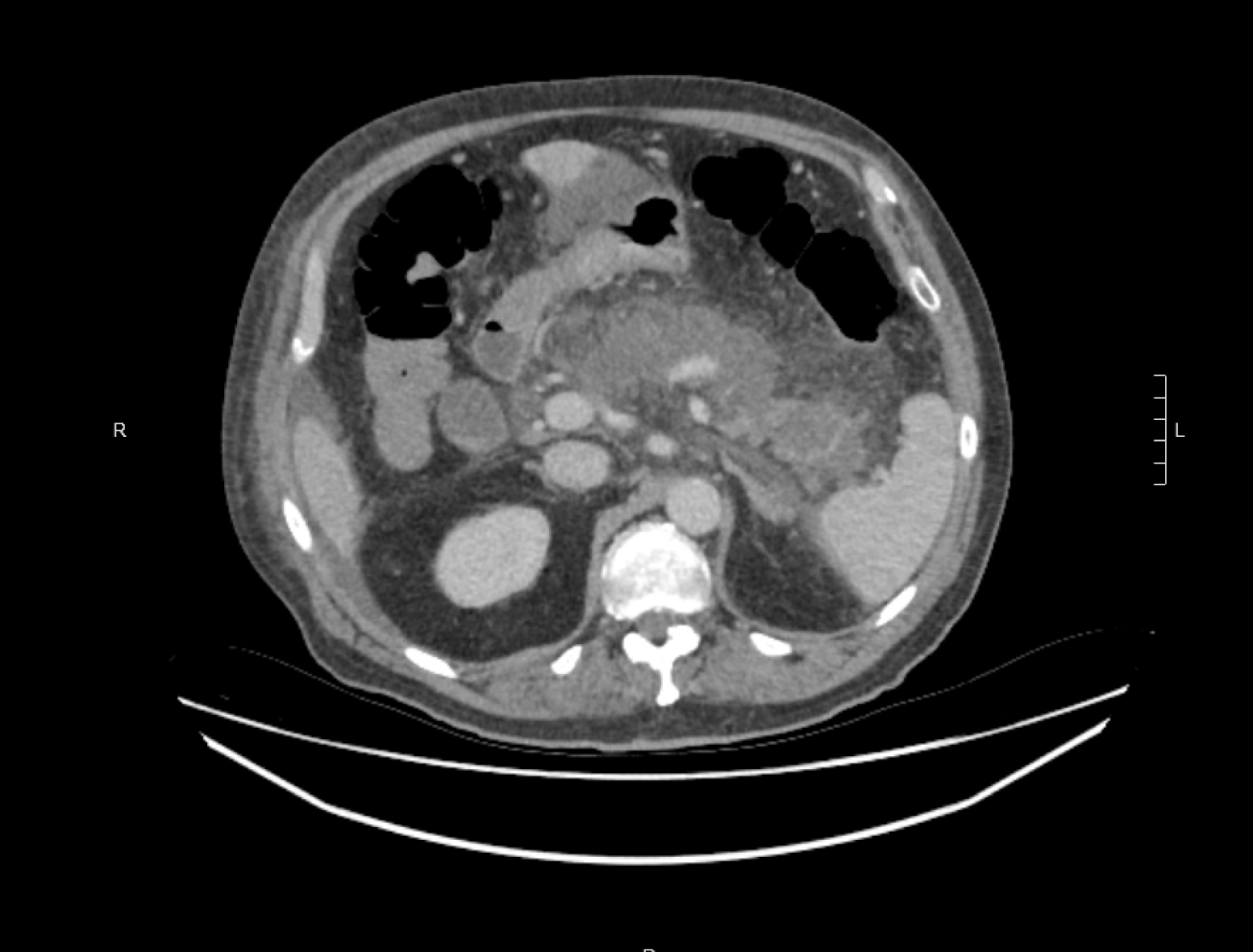
Figure 1 Axial contrast enhanced computed tomography scan showing extensive pancreatic necrosis with non-enhancement of the pancreas.
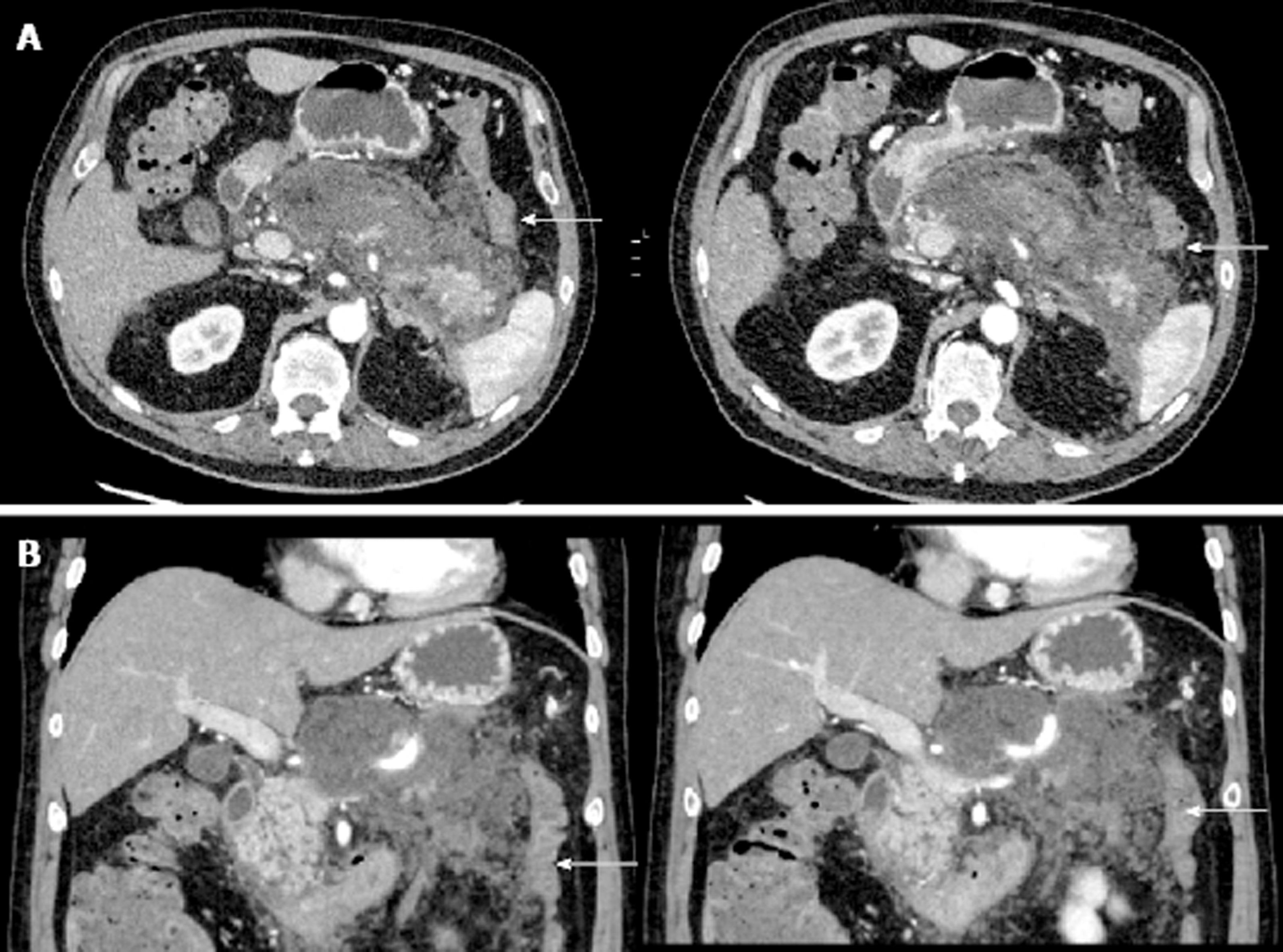
Figure 2 Contrast enhanced computed tomography scan showing peripancreatic oedema and mesocolic inflammatory change.
A: Axial contrast enhanced computed tomography (CT) scan showing marked peripancreatic oedema extending into the lesser sac and to the splenic flexure (arrow showing splenic flexure of colon); B: Coronal contrast enhanced CT scan showing colon (arrow) with adjacent mesocolic inflammatory change secondary to pancreatitis.
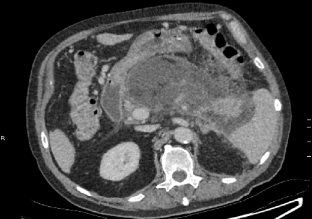
Figure 3 Axial contrast enhanced computed tomography scan showing interval increase in size and peripheral enhancement of walled of pancreatic necrosis.
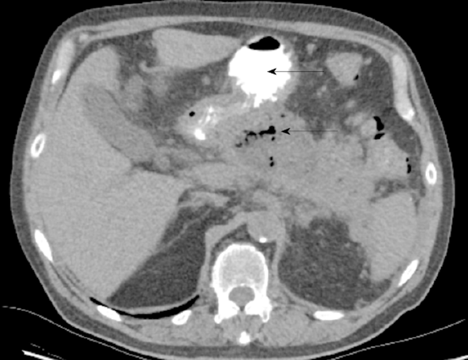
Figure 4 Walled of necrosis with locules of gas.
Oral contrast seen within the stomach (solid arrow).
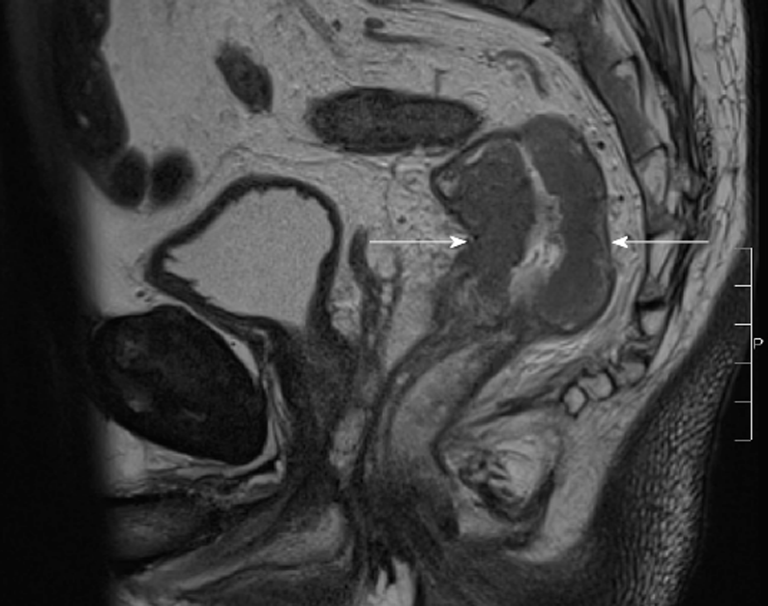
Figure 5 T2 sagittal view of magnetic resonance imaging rectum.
Arrows showing rectal cancer.
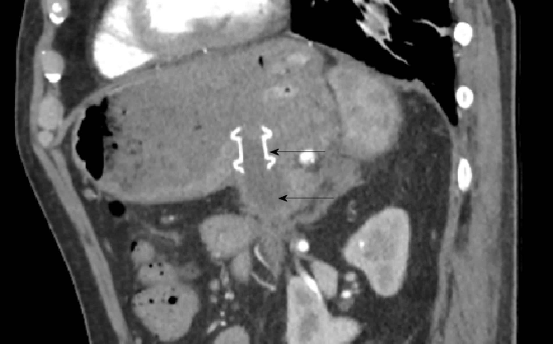
Figure 6 Sagittal computed tomography exam.
AXIOS cystogastrostomy stent Solid arrow showing stent. Hollow arrow delineating small residual cyst.
- Citation: Choi K, Flynn DE, Karunairajah A, Hughes A, Bhasin A, Devereaux B, Chandrasegaram MD. Management of infected pancreatic necrosis in the setting of concomitant rectal cancer: A case report and review of literature. World J Gastrointest Surg 2019; 11(4): 237-246
- URL: https://www.wjgnet.com/1948-9366/full/v11/i4/237.htm
- DOI: https://dx.doi.org/10.4240/wjgs.v11.i4.237


