
















Copyright: ©Author(s) 2026.
World J Diabetes. Oct 15, 2026; 17(10): 124847
Published online Oct 15, 2026. doi: 10.4239/wjd.124847
Published online Oct 15, 2026. doi: 10.4239/wjd.124847
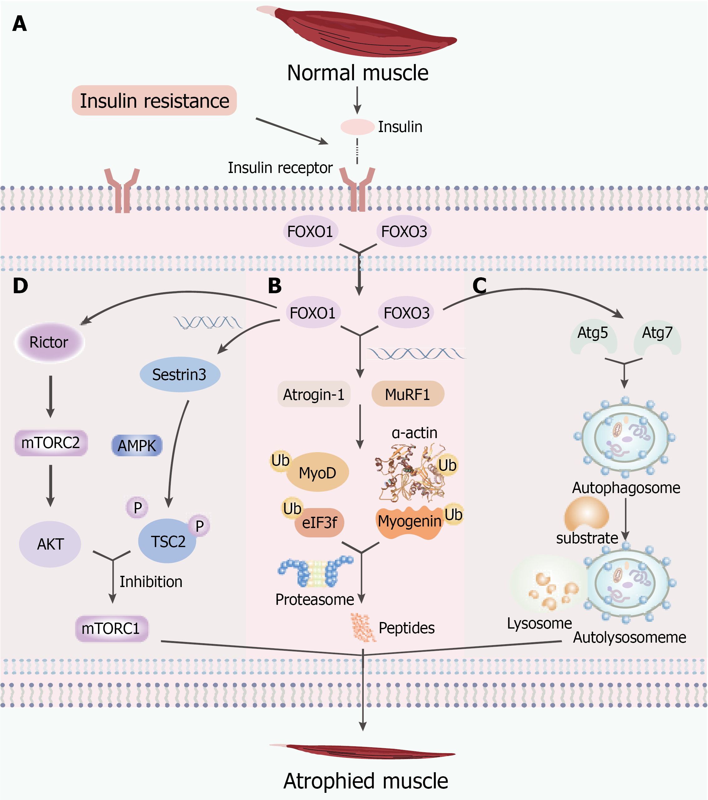
Figure 1 Mechanisms of insulin resistance-induced skeletal muscle atrophy.
A: Insulin resistance impairs insulin receptor-mediated phosphoinositide 3-kinase-protein kinase B (AKT) signaling, resulting in reduced AKT activity and subsequent nuclear translocation of forkhead box O transcription factors (FOXO) 1 and FOXO3; B: Activated FOXO1/FOXO3 promote the transcription of muscle-specific ubiquitin ligases, including Atrogin-1 and MuRF1, which induce ubiquitination and proteasomal degradation of myogenic regulatory proteins (MyoD, myogenin, and eIF3f) and contractile proteins (α-actin), thereby accelerating muscle protein degradation; C: FOXO3 activates autophagy-related genes, including Atg5 and Atg7, promoting autophagosome formation and autolysosomal degradation, which further contributes to skeletal muscle wasting; D: FOXO1 inhibits mechanistic target of rapamycin complex 1-mediated protein synthesis through both tuberous sclerosis complex 2 (TSC2)-dependent and TSC2-independent pathways. The Sestrin3-AMP-activated protein kinase-TSC2 pathway and rapamycin-insensitive companion of mechanistic target of rapamycin-mechanistic target of rapamycin complex 2-AKT pathway suppress mechanistic target of rapamycin complex 1 activity, leading to impaired muscle protein synthesis. AKT: Protein kinase B; FOXO: Forkhead box O transcription factors; mTORC1: Mechanistic target of rapamycin complex 1; mTORC2: Mechanistic target of rapamycin complex 2; TSC2: Tuberous sclerosis complex 2; Rictor: Rapamycin-insensitive companion of mechanistic target of rapamycin; AMPK: AMP-activated protein kinase.
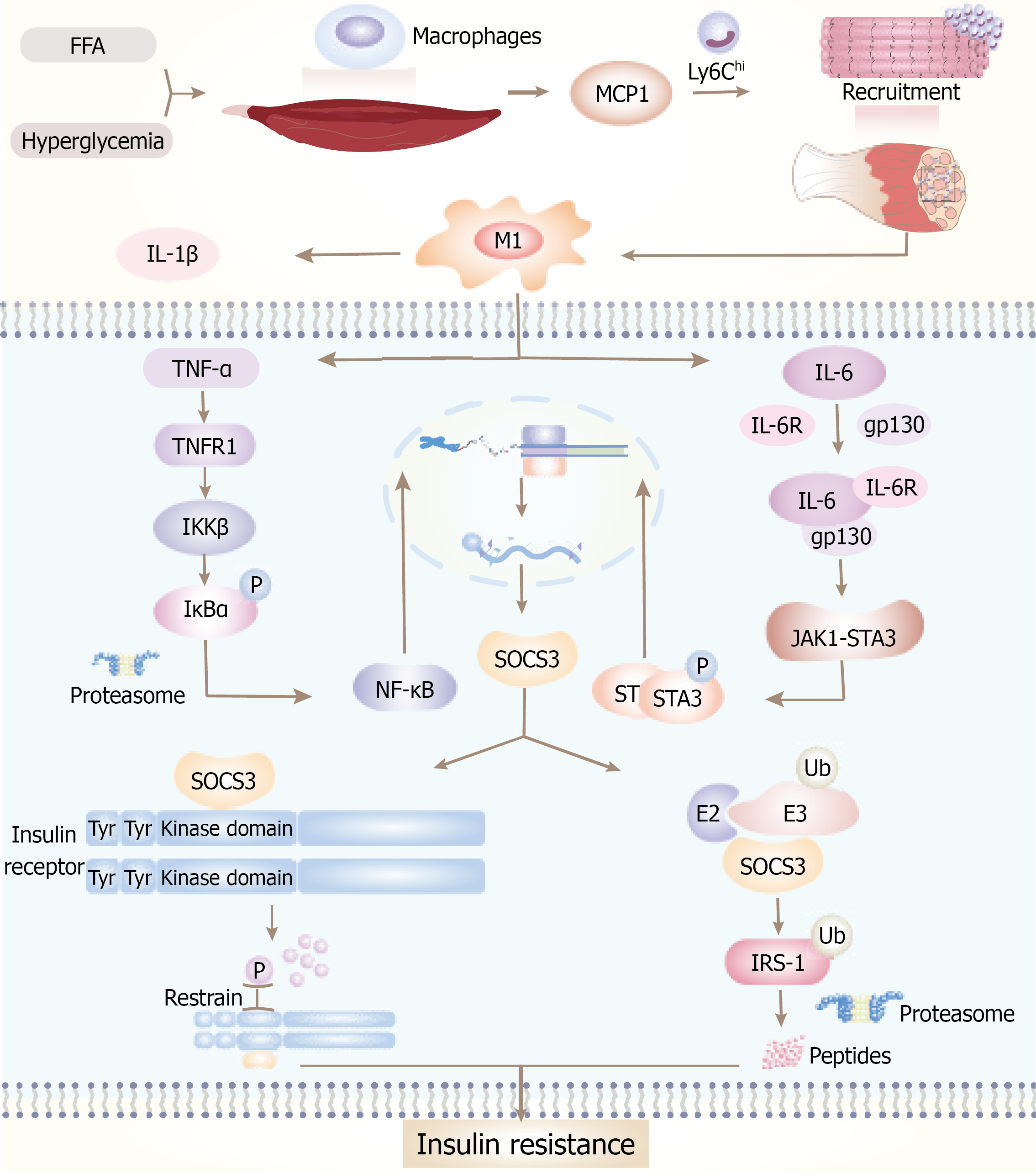
Figure 2 Inflammation induces insulin resistance in skeletal muscle.
Elevated levels of glucose and free fatty acid stimulate the secretion of monocyte chemoattractant protein-1 from both muscle fibers and resident macrophages. This chemokine facilitates the recruitment of Ly6Chi monocytes, which subsequently differentiate into pro-inflammatory M1 macrophages. These macrophages secrete pro-inflammatory cytokines, including tumor necrosis factor-alpha, interleukin (IL)-6, and IL-1β. tumor necrosis factor-alpha activates the IKKβ-IκBα-nuclear factor kappa-B signaling cascade, whereas IL-6 induces phosphorylation of the JAK1-STAT3 pathway. The convergence of these signaling pathways leads to the upregulation of suppressor of cytokine signaling 3, which impairs insulin signaling by inhibiting insulin receptor autophosphorylation and promoting the ubiquitination and subsequent degradation of insulin receptor substrate-1. Collectively, these molecular events culminate in the development of insulin resistance within skeletal muscle tissue. MCP-1: Monocyte chemoattractant protein-1; TNF-α: Tumor necrosis factor-alpha; IL-6: Interleukin-6; SOCS3: Suppressor of cytokine signaling 3; FFA: Free fatty acid; IRS-1: Insulin receptor substrate-1.
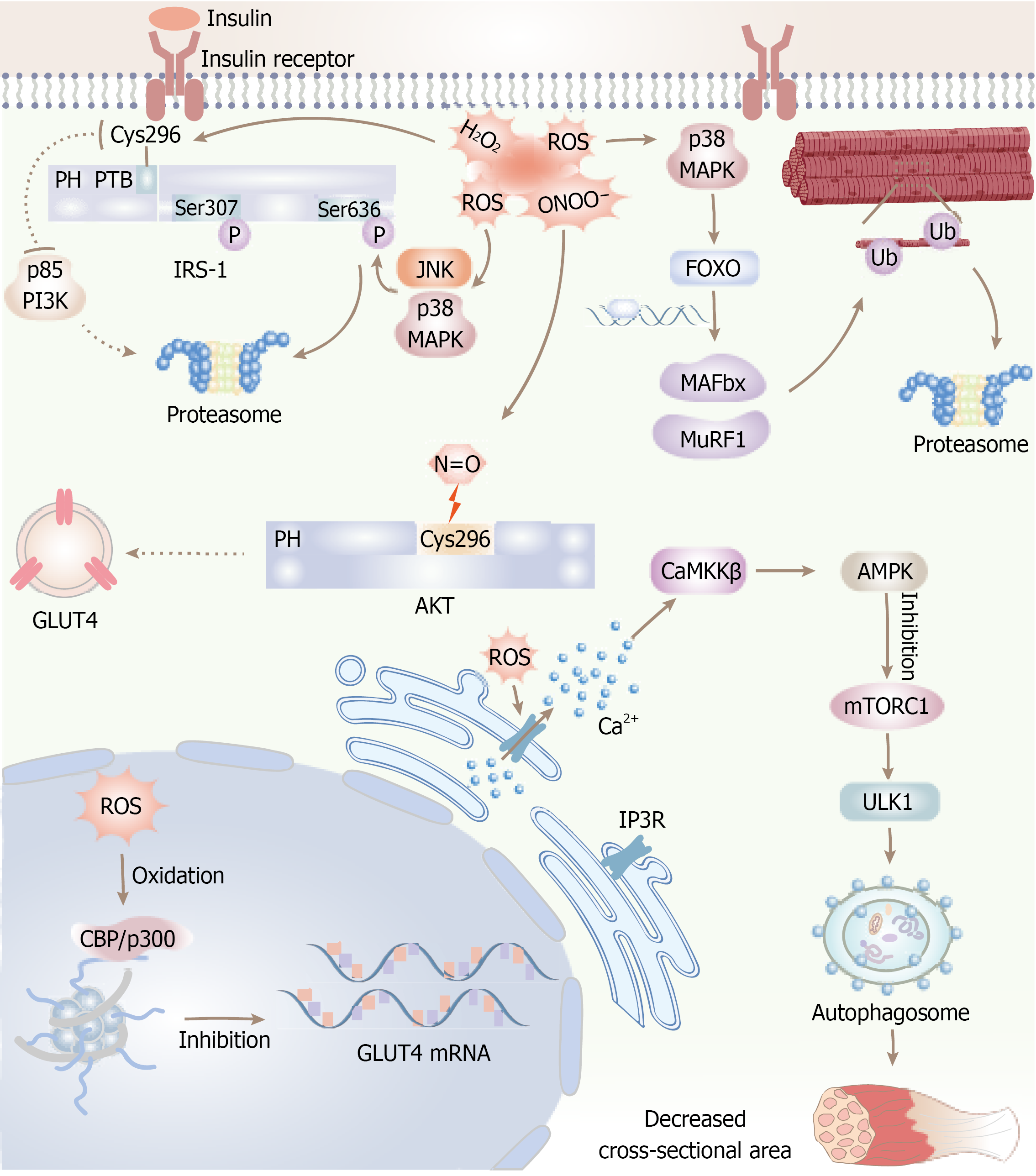
Figure 3 Reactive oxygen species contribute to insulin resistance and muscle atrophy in skeletal muscle through several molecular mechanisms.
Specifically, hydrogen peroxide (H2O2) directly oxidizes cysteine residue 296 on insulin receptor substrate-1, thereby inhibiting the recruitment of phosphoinositide 3-kinase. Reactive oxygen species (ROS)-mediated activation of JNK and p38 MAPK leads to the inhibitory phosphorylation of insulin receptor substrate-1 at serine residues 307 and 636, which facilitates its ubiquitination and subsequent proteasomal degradation. Peroxynitrite (ONOO-) nitrates cysteine 296 on protein kinase B, resulting in diminished kinase activity and impaired translocation of GLUT4. Furthermore, ROS induces oxidation of the transcriptional coactivators CBP/p300, causing epigenetic repression of GLUT4 gene expression. Concurrently, ROS activates the p38 MAPK- forkhead box O transcription factors signaling axis, which upregulates the expression of muscle-specific E3 ubiquitin ligases MAFbx and MuRF1, thereby promoting proteasomal protein degradation. Moreover, ROS triggers calcium release through oxidation of IP3R, which activates the Ca2+/CaMKKβ/AMP-AMP-activated protein kinase pathway. This activation inhibits mechanistic target of rapamycin complex 1 and stimulates ULK1-mediated autophagy, collectively contributing to a reduction in muscle fiber cross-sectional area. ROS: Reactive oxygen species; IRS-1: Insulin receptor substrate-1; PI3K: Phosphoinositide 3-kinase; AKT: Protein kinase B; FOXO: Forkhead box O transcription factors; mTORC1: Mechanistic target of rapamycin complex 1.
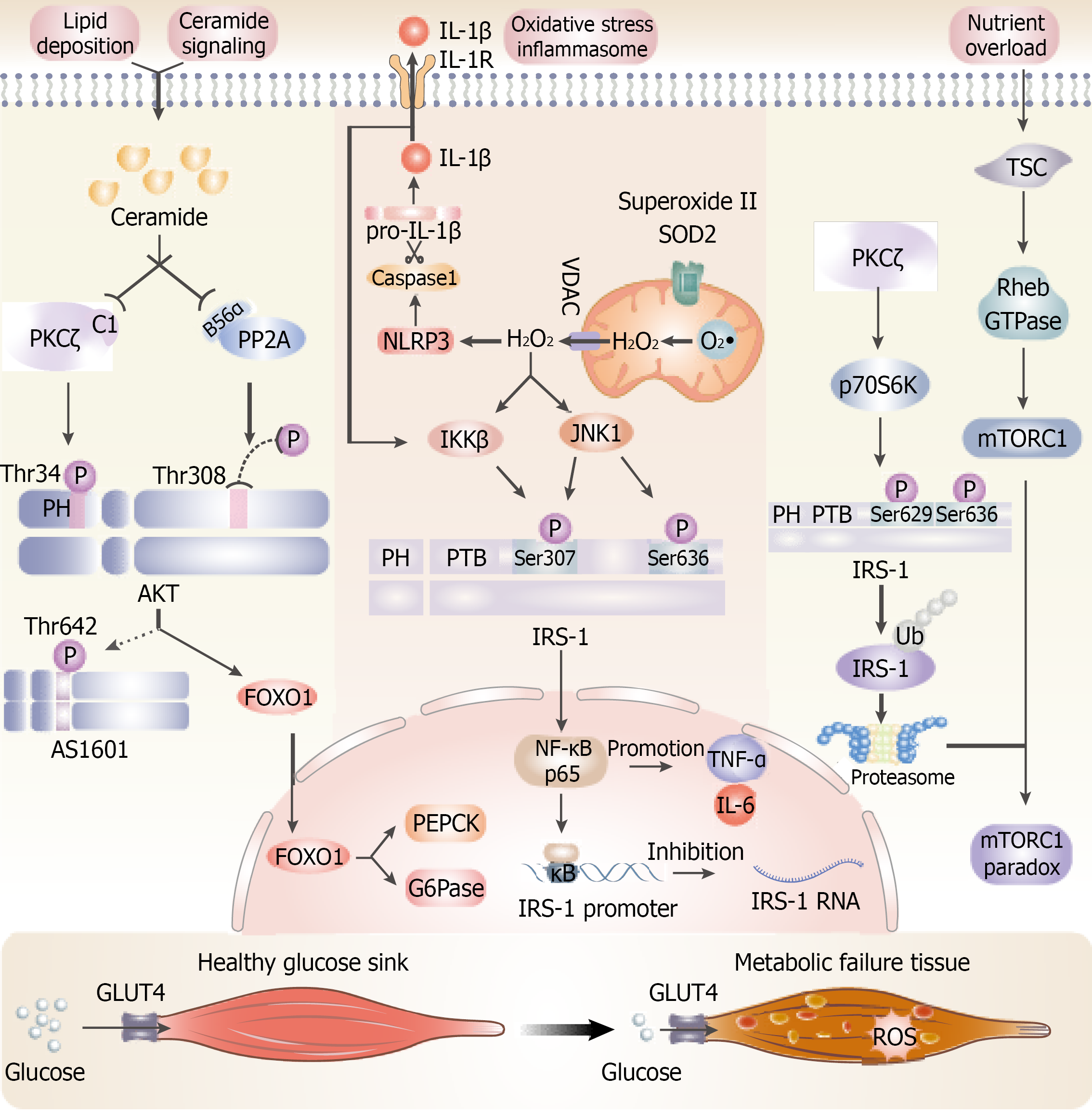
Figure 4 Mechanisms underlying ceramide-induced and reactive oxygen species-induced insulin resistance in skeletal muscle involve multiple interconnected pathways.
Ceramide impairs protein kinase B activity through inhibitory phosphorylation mediated by PKCζ and dephosphorylation facilitated by PP2A, thereby disrupting GLUT4 translocation. Concurrently, mitochondrial hydrogen peroxide (H2O2) activates IKKβ and JNK1, which promote inhibitory phosphorylation and subsequent degradation of insulin receptor substrate-1 (IRS-1). nuclear factor kappa-B suppresses IRS-1 gene transcription. Reactive oxygen species further stimulate the NLRP3 inflammasome, leading to interleukin-1β production that perpetuates inflammatory signaling. Nutrient excess activates the mechanistic target of rapamycin complex 1. PKCζ-mediated activation of p70S6K induces feedback phosphorylation of IRS-1. These coordinated molecular events convert skeletal muscle from an efficient glucose-utilizing tissue into one characterized by chronic insulin resistance and metabolic dysfunction. TNF-α: Tumor necrosis factor-alpha; IL: Interleukin; AKT: Protein kinase B; FOXO: Forkhead box O transcription factors; SOD: Superoxide dismutase; IRS-1: Insulin receptor substrate-1; TSC: Tuberous sclerosis complex; mTORC1: Mechanistic target of rapamycin complex 1.
- Citation: Xie XL, Cao HY, Wang ZT, Chen HR, Zhou LS, Qian T, Han ZY. Pathophysiological mechanisms and risk factors linking type 2 diabetes mellitus and sarcopenia: A narrative review. World J Diabetes 2026; 17(10): 124847
- URL: https://www.wjgnet.com/1948-9358/full/v17/i10/124847.htm
- DOI: https://dx.doi.org/10.4239/wjd.124847