Published online Aug 16, 2023. doi: 10.4253/wjge.v15.i8.540
Peer-review started: January 4, 2023
First decision: April 13, 2023
Revised: April 18, 2023
Accepted: July 17, 2023
Article in press: July 17, 2023
Published online: August 16, 2023
Processing time: 217 Days and 11.3 Hours
Endoscopic biopsy is mandatory for the diagnosis of malignant and premalignant ampullary tumours. The commonly reported inadvertent complications following routine mucosal biopsy include perforation and haemorrhage. Acute pancreatitis is an extremely rare complication following this procedure.
This report details the case of a 59-year-old man who underwent biopsy of the ampulla for a suspected periampullary tumour. Following the procedure, the patient presented with symptoms of acute pancreatitis which was substantiated by laboratory and radiological investigations. He was conservatively managed and discharged following complete resolution of symptoms.
This case report serves to highlight the importance of this potential complication following routine endoscopic biopsy of the ampulla.
Core Tip: Gastrointestinal endoscopic procedures are relatively safe and are being routinely performed with the advent of minimally invasive procedures. Acute pancreatitis is an extremely uncommon complication following endoscopic ampullary biopsy. It is important for endoscopists to be mindful of this untoward complication with appropriate post-procedure monitoring and support.
- Citation: George NM, Rajesh NA, Chitrambalam TG. Acute pancreatitis following endoscopic ampullary biopsy: A case report. World J Gastrointest Endosc 2023; 15(8): 540-544
- URL: https://www.wjgnet.com/1948-5190/full/v15/i8/540.htm
- DOI: https://dx.doi.org/10.4253/wjge.v15.i8.540
Endoscopic biopsy is recommended for the evaluation of ampullary adenomas, ampullary tumours, and more recently, immunohistological staining for autoimmune pancreatitis[1,2]. The commonly encountered complications following this procedure include bleeding, infection, and perforation. Acute pancreatitis is an extremely uncommon complication with a high rate of morbidity and mortality. It can be attributed to the mucosal edema or intraductal hematoma caused by the ampullary biopsy[6]. Although rare, endoscopists are to be aware of this complication and patients need to be closely monitored following the procedure.
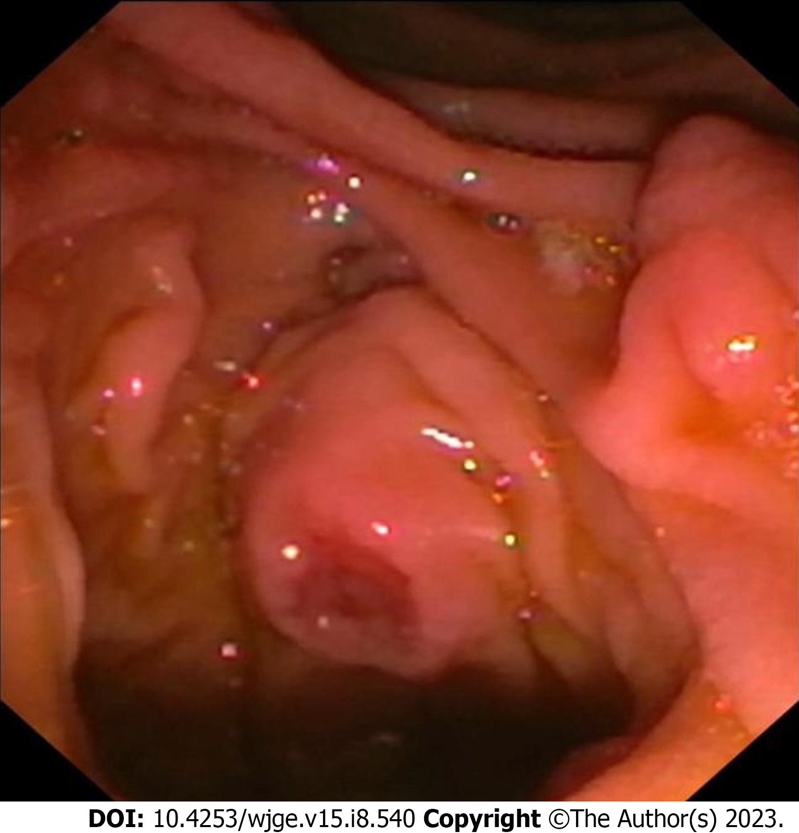
A 59-year-old man presented to our tertiary centre with symptoms of dyspepsia for which ultrasound of the abdomen was done and it showed dilatation of the common bile duct (10 mm). For further evaluation, liver function test was done, which was reported as normal. Contrast-enhanced computed tomography (CT) of the abdomen was then performed, which revealed dilatation of the common bile duct and pancreatic duct (3.5 mm). Side-viewing duodenoscopy (Olympus TJF-150 Video Duodenoscope; Olympus, Tokyo, Japan) was done, which revealed an ulcerated papilla from which a biopsy was taken (Figure 1). The sampling was done with Jumbo biopsy forceps without spike. Haemostasis was confirmed and the procedure was uneventful. Two hours later, the patient presented with acute onset upper abdominal pain and profuse sweating which developed 30 min following his meal.
The pain was localised to the epigastrium and was severe in nature (8 on the Visual Analogue Scale) with radiation to the back. There was no history of vomiting.
The patient was not a known diabetic or hypertensive.
The patient did not have any relevant family history. He was a non-alcoholic and non-smoker.
At the Emergency Room, the patient’s heart rate was 110 per minute and blood pressure was 140/80 mm of Hg. On examination of the abdomen, there was severe epigastric tenderness with guarding. The rest of the abdominal quadrants were non-tender with normal bowel sounds.
The patient’s blood work-up pre- and post-procedure is shown in Table 1.
| Blood investigation | Pre-procedure | Post-procedure |
| WBC count | 7500/mm3 | 13000/mm3 |
| AST | 35 IU/L | 65 IU/L |
| ALT | 40 IU/L | 82 IU/L |
| Serum amylase | 50 IU/L | 1500 IU/L |
| Serum lipase | 110 IU/L | 800 IU/L |
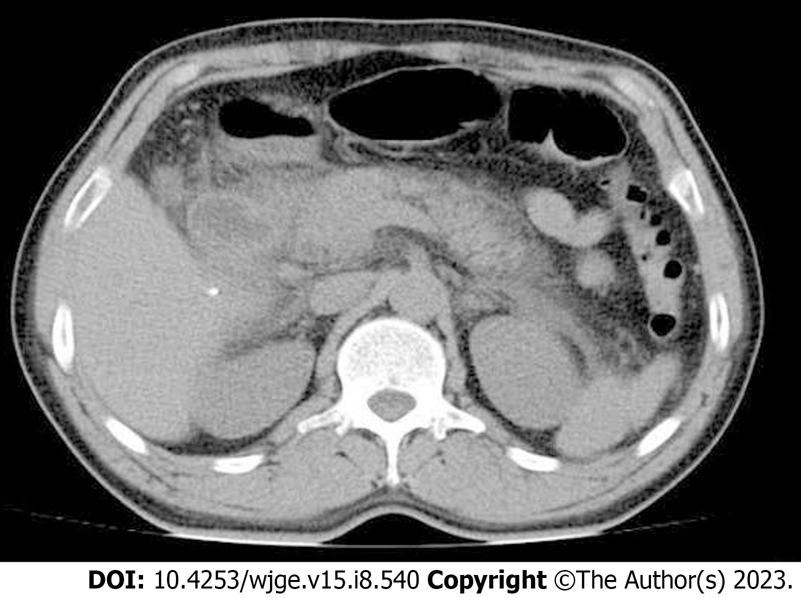
Computed tomography of the abdomen showed features consistent with acute pancreatitis such as pancreatic enlargement and diffuse peri-pancreatic fat stranding (Figure 2).
The patient was further evaluated to determine other attributing factors causing pancreatitis such as gallstone disease, alcohol, or any other precipitating drugs. After ruling these out, endoscopic biopsy of the ampulla was attributed as the cause.
The patient was admitted and kept nil per oral. He was managed conservatively with intravenous fluids, antibiotics, and analgesics. His general condition improved and he was gradually initiated on diet. He achieved complete resolution of symptoms and was discharged 48 h later.
Histopathological examination of the tissues samples showed an adenomatous polyp with moderate dysplasia. The patient remained asymptomatic over a follow-up period of 6 mo.
Upper gastrointestinal endoscopy is central for the diagnosis of a wide array of tumours arising at the ampulla of Vater including neoplasms such as neuroendocrine tumours, adenomas, and adenocarcinomas as well as non-neoplastic lesions such as lipomas, lymphangiomas, fibromas, adenomyomas, and hamartomas[3-5]. Acute pancreatitis, a commonly encountered complication following endoscopic retrograde cholangiopancreatography, is extremely rare following non-thermal endoscopic biopsy of the ampulla of Vater without previous cannulation. Morales et al[6], who reported the first such case in 1994, propositioned mucosal edema or intraductal hematoma with a resultant increase in pressure in the pancreatic duct as the cause. Ishida et al[7] presented a similar case of acute pancreatitis following endoscopic biopsy of the ampulla of Vater in 2013, where the cause was ascribed to the small ampulla of the patient. Confirmation of hemostasis at the end of the procedure is important in order to prevent the inadvertent development of acute pancreatitis as a result of intramural hematoma. Another contributing factor is the ampullary edema as a result of the biopsy forceps. Ampullary biopsy with side-viewing endoscopy is pivotal for the diagnosis of periampullary carcinoma. However, the yield of ampullary surface biopsies is limited and there arises the need for deeper biopsies which can further contribute to ampullary edema. In a case of acute pancreatitis following endoscopic ampullary biopsy reported by Michopoulos et al[8], they directed the biopsies to the area around the orifice. It is recommended to avoid biopsying the normal ampulla and to biopsy some distance from the mouth of the pancreatic duct to prevent acute pancreatitis; however, bleeding and edema can obscure vision, proving this to be difficult. There are very limited reported cases of acute pancreatitis following endoscopic biopsies from the ampulla of Vater. Most of these patients have had an uneventful recovery. Skelton et al[9] reported a case of severe necrotising pancreatitis following ampullary biopsy where the patient required multiple necrosectomies and two CT-guided drains. In our case, the patient was discharged 48 h post-procedure without any untoward outcomes.
This case reports serves to enlighten endoscopists regarding the potential complication of acute pancreatitis following endoscopic biopsy of the ampulla, to educate patients regarding this complication, and to closely monitor them following the procedure.
| 1. | Tsukada K, Takada T, Miyazaki M, Miyakawa S, Nagino M, Kondo S, Furuse J, Saito H, Tsuyuguchi T, Kimura F, Yoshitomi H, Nozawa S, Yoshida M, Wada K, Amano H, Miura F; Japanese Association of Biliary Surgery; Japanese Society of Hepato-Biliary-Pancreatic Surgery; Japan Society of Clinical Oncology. Diagnosis of biliary tract and ampullary carcinomas. J Hepatobiliary Pancreat Surg. 2008;15:31-40. [RCA] [PubMed] [DOI] [Full Text] [Full Text (PDF)] [Cited by in Crossref: 30] [Cited by in RCA: 36] [Article Influence: 2.0] [Reference Citation Analysis (0)] |
| 2. | Kamisawa T, Tu Y, Egawa N, Tsuruta K, Okamoto A. A new diagnostic endoscopic tool for autoimmune pancreatitis. Gastrointest Endosc. 2008;68:358-361. [RCA] [PubMed] [DOI] [Full Text] [Cited by in Crossref: 71] [Cited by in RCA: 61] [Article Influence: 3.4] [Reference Citation Analysis (0)] |
| 3. | Hartel M, Wente MN, Sido B, Friess H, Büchler MW. Carcinoid of the ampulla of Vater. J Gastroenterol Hepatol. 2005;20:676-681. [RCA] [PubMed] [DOI] [Full Text] [Cited by in Crossref: 89] [Cited by in RCA: 71] [Article Influence: 3.4] [Reference Citation Analysis (0)] |
| 4. | Cho YS, Joo HJ, Seo EK, Jeon WJ, Chae HB, Park SM, Sung RH. A case of juxtapapillary gangliocytic paraganglioma treated with endoscopic resection. Korean J Med. 2010;79:543-548 Available from:https://www.koreamed.org/SearchBasic.php?RID=1458432. |
| 5. | Artaza T, Potenciano JM, Legaz M, Muñoz C, Talavera A, Sánchez E. Lymphangioma of Vater's ampulla: a rare cause of obstructive jaundice. Endoscopic therapy. Scand J Gastroenterol. 1995;30:804-806. [RCA] [PubMed] [DOI] [Full Text] [Cited by in Crossref: 5] [Cited by in RCA: 4] [Article Influence: 0.1] [Reference Citation Analysis (0)] |
| 6. | Morales TG, Hixson LJ. Acute pancreatitis following endoscopic biopsy of the ampulla in a patient with Gardner's syndrome. Gastrointest Endosc. 1994;40:367-369. [RCA] [PubMed] [DOI] [Full Text] [Cited by in Crossref: 10] [Cited by in RCA: 11] [Article Influence: 0.3] [Reference Citation Analysis (0)] |
| 7. | Ishida Y, Okabe Y, Tokuyasu H, Kaji R, Sugiyama G, Ushijima T, Sasaki Y, Yasumoto M, Kuraoka K, Tsuruta O, Sata M. A case of acute pancreatitis following endoscopic biopsy of the ampulla of Vater. Kurume Med J. 2013;60:67-70. [RCA] [PubMed] [DOI] [Full Text] [Cited by in Crossref: 8] [Cited by in RCA: 12] [Article Influence: 1.0] [Reference Citation Analysis (0)] |
| 8. | Michopoulos S, Kozompoli D, Ntai S, Kalantzis G, Zampeli E, Petraki K. Acute Pancreatitis Following Endoscopic Ampullary Biopsies without Attempted Cannulation of the Ampulla of Vater. Clin Endosc. 2016;49:575-578. [RCA] [PubMed] [DOI] [Full Text] [Full Text (PDF)] [Cited by in Crossref: 2] [Cited by in RCA: 6] [Article Influence: 0.6] [Reference Citation Analysis (0)] |
| 9. | Skelton D, Barnes J, French J. A case of severe necrotising pancreatitis following ampullary biopsy. Ann R Coll Surg Engl. 2015;97:e61-e63. [RCA] [PubMed] [DOI] [Full Text] [Cited by in Crossref: 6] [Cited by in RCA: 4] [Article Influence: 0.4] [Reference Citation Analysis (0)] |
Open-Access: This article is an open-access article that was selected by an in-house editor and fully peer-reviewed by external reviewers. It is distributed in accordance with the Creative Commons Attribution NonCommercial (CC BY-NC 4.0) license, which permits others to distribute, remix, adapt, build upon this work non-commercially, and license their derivative works on different terms, provided the original work is properly cited and the use is non-commercial. See: https://creativecommons.org/Licenses/by-nc/4.0/
Provenance and peer review: Unsolicited article; Externally peer reviewed.
Peer-review model: Single blind
Specialty type: Gastroenterology and hepatology
Country/Territory of origin: India
Peer-review report’s scientific quality classification
Grade A (Excellent): 0
Grade B (Very good): 0
Grade C (Good): C
Grade D (Fair): D
Grade E (Poor): 0
P-Reviewer: Askarpour H, Japan; Kitamura K, Japan S-Editor: Liu JH L-Editor: Wang TQ P-Editor: Cai YX


