©The Author(s) 2017.
World J Gastrointest Endosc. Sep 16, 2017; 9(9): 486-493
Published online Sep 16, 2017. doi: 10.4253/wjge.v9.i9.486
Published online Sep 16, 2017. doi: 10.4253/wjge.v9.i9.486
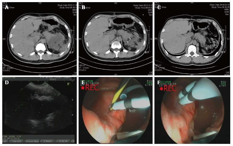
Figure 1 Endoscopic ultrasound-guided drainage of pancreatic fluid collection.
A: CT scan before procedure; B: CT scan after procedure (24 h); C: CT scan after procedure (1 mo); D: EUS imaging of PFC; E and F: endoscopic imaging of double pig-tail tubes implantation. EUS: Endoscopic ultrasound; PFC: Pancreatic fluid collection; CT: Computed tomography.
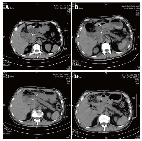
Figure 2 Re-intervention of pancreatic fluid collection drainage.
A: Computed tomography (CT) scan before procedure; B: CT scan after first procedure (72 h); C: CT scan after re-intervention (72 h); D: CT scan after procedure (1 mo).
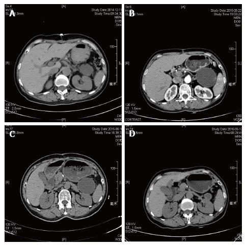
Figure 3 Failed case of pancreatic fluid collection drainage.
A: Computed tomography (CT) scan after procedure (24 h); B: CT scan after procedure (72 h); C: CT scan after procedure (3 mo); D: CT scan after procedure (1 year).
- Citation: Hao SJ, Xu WJ, Di Y, Yao L, He H, Yang F, Jin C, Zhong L, Fu DL. Novel and supplementary management of pancreatic fluid collections: Endoscopic ultrasound-guided drainage. World J Gastrointest Endosc 2017; 9(9): 486-493
- URL: https://www.wjgnet.com/1948-5190/full/v9/i9/486.htm
- DOI: https://dx.doi.org/10.4253/wjge.v9.i9.486


