Copyright
©The Author(s) 2017.
World J Gastrointest Endosc. Jul 16, 2017; 9(7): 327-333
Published online Jul 16, 2017. doi: 10.4253/wjge.v9.i7.327
Published online Jul 16, 2017. doi: 10.4253/wjge.v9.i7.327
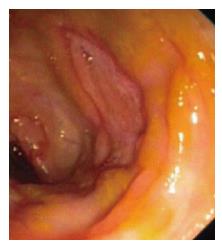
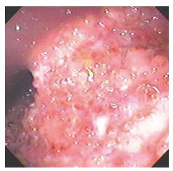
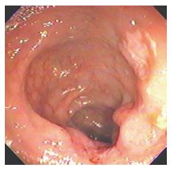
Figure 1 Amoebic ulcer in cecum.
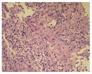
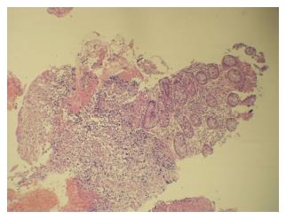
Figure 2 Amoebic trophozoites on histopathology.
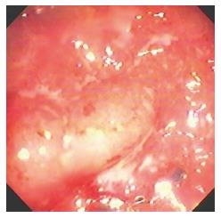
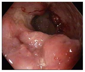
Figure 3 Tubercular ulcers in ileum.
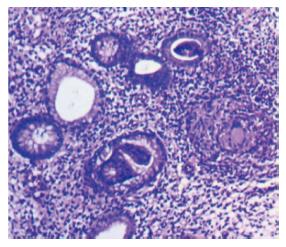
Figure 4 Tubercular granuloma.
Figure 5 Pseudopolyps in Crohn’s disease.
Figure 6 Ulcer on ileo-cecal valve ileocecal-lymphoma.
Figure 7 Ileal ulcer-lymphoma.
Figure 8 Biopsy demonstrating lymphoma.
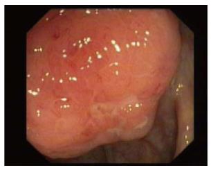
Figure 9 Non-specific ileal ulcer.
- Citation: Toshniwal J, Chawlani R, Thawrani A, Sharma R, Arora A, Kotecha HL, Goyal M, Kirnake V, Jain P, Tyagi P, Bansal N, Sachdeva M, Ranjan P, Kumar M, Sharma P, Singla V, Bansal R, Shah V, Bhalla S, Kumar A. All ileo-cecal ulcers are not Crohn’s: Changing perspectives of symptomatic ileocecal ulcers. World J Gastrointest Endosc 2017; 9(7): 327-333
- URL: https://www.wjgnet.com/1948-5190/full/v9/i7/327.htm
- DOI: https://dx.doi.org/10.4253/wjge.v9.i7.327


