©The Author(s) 2017.
World J Gastrointest Endosc. Dec 16, 2017; 9(12): 583-589
Published online Dec 16, 2017. doi: 10.4253/wjge.v9.i12.583
Published online Dec 16, 2017. doi: 10.4253/wjge.v9.i12.583
Figure 1 Abdominal dynamic contrast-enhanced computed tomography showed a 27-mm diameter tumor in the pancreatic uncus, which was well defined and enhanced from the arterial phase, exhibiting the greatest enhancement in the arterial phase.
White arrow indicates the tumor.
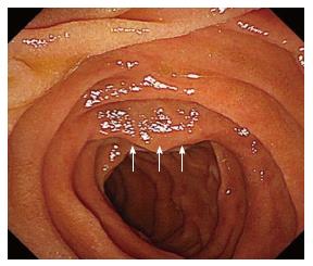
Figure 2 A slightly elevated lesion located in the inferior angle of the duodenum with normal overlying mucosa was detected on upper gastrointestinal endoscopy.
White arrows indicate the elevation.
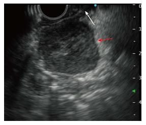
Figure 3 Endoscopic ultrasonography showed a well-defined hypoechoic mass in the pancreatic uncus, and the tumor connected with the muscularis propria layer of the duodenum.
Red arrow indicates the tumor and white arrow indicates the muscularis propria layer.
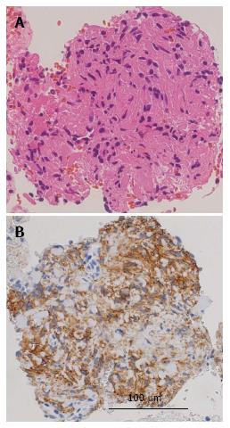
Figure 4 Histological analysis of specimen collected by endoscopic ultrasound-guided fine-needle aspiration.
A: Hematoxylin and eosin staining revealed that the tumor was mainly composed of spindle-shaped cells; B: The tumor cells were positive for c-kit.
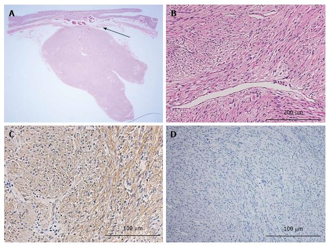
Figure 5 Histological analysis of resected tumor tissue.
A: Macroscopic finding showed 30 mm × 22 mm × 22 mm sized tumor showing extraluminal growth from duodenum (black arrow); B: Hematoxylin and eosin-stained sections showed that the tumor was mainly composed of spindle-shaped cells without necrosis; C: The tumor cells appeared immunohistochemically positive for c-kit; D: Mitosis was detected in 2/50 high-power fields, and MIB-1 labeling index (Ki-67 stain) was < 1%.
- Citation: Hayashi K, Kamimura K, Hosaka K, Ikarashi S, Kohisa J, Takahashi K, Tominaga K, Mizuno K, Hashimoto S, Yokoyama J, Yamagiwa S, Takizawa K, Wakai T, Umezu H, Terai S. Endoscopic ultrasound-guided fine-needle aspiration for diagnosing a rare extraluminal duodenal gastrointestinal tumor. World J Gastrointest Endosc 2017; 9(12): 583-589
- URL: https://www.wjgnet.com/1948-5190/full/v9/i12/583.htm
- DOI: https://dx.doi.org/10.4253/wjge.v9.i12.583


