©The Author(s) 2017.
World J Gastrointest Endosc. Oct 16, 2017; 9(10): 529-534
Published online Oct 16, 2017. doi: 10.4253/wjge.v9.i10.529
Published online Oct 16, 2017. doi: 10.4253/wjge.v9.i10.529
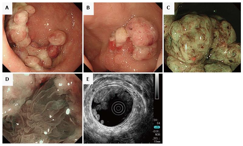
Figure 1 Endoscopic findings of multiple inflammatory polyps.
A and B: Caps of fibrinopurulent exudate from the sigmoid colon to the rectum interspersed with normal colonic mucosa; C and D: Magnifying endoscopy shows an area in the cap of amorphous fibrinopurulent exudate and tortuous and long branching crypts under the cap; E: Endoscopic ultrasonography showed significant thickening of the colonic mucosa layers.
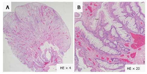
Figure 2 Microscopic findings of inflammatory polyps show elongated, tortuous, branched, and dilated crypts with epithelial hyperplasia, inflammatory granulation tissue and a mild degree of fibromusculosis in the lamina propria.
Hematoxylin-Eosin stain, × 4 (A) and × 20 (B).
Figure 3 Multiple elevated lesions with wall thickening in the sigmoid colon.
Rectum on computed tomography (A) and magnetic resonance imaging (B); Barium enema shows a collection of small sessile polyps in the sigmoid colon and rectum (C).
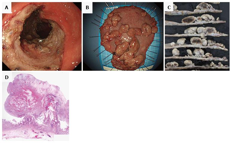
Figure 4 Endoscopic submucosal dissection.
Post-dissection ulcers after endoscopic submucosal dissection at the sigmoid colon and rectum (A). Fresh specimen of cap polyposis after endoscopic submucosal dissection (B) and fixed specimen (C). The fixed specimen revealed bleeding into the polyps. Microscopic findings of inflammatory polyps [HE stain, × 4 (D)].
- Citation: Murata M, Sugimoto M, Ban H, Otsuka T, Nakata T, Fukuda M, Inatomi O, Bamba S, Kushima R, Andoh A. Cap polyposis refractory to Helicobacter pylori eradication treated with endoscopic submucosal dissection. World J Gastrointest Endosc 2017; 9(10): 529-534
- URL: https://www.wjgnet.com/1948-5190/full/v9/i10/529.htm
- DOI: https://dx.doi.org/10.4253/wjge.v9.i10.529


