Copyright
©The Author(s) 2016.
World J Gastrointest Endosc. Apr 10, 2016; 8(7): 319-329
Published online Apr 10, 2016. doi: 10.4253/wjge.v8.i7.319
Published online Apr 10, 2016. doi: 10.4253/wjge.v8.i7.319
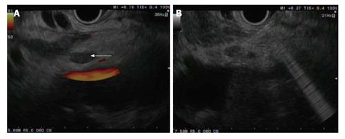
Figure 1 Endoscopic ultrasound-guided injection into the celiac ganglion.
A: Celiac ganglion visualized by linear endoscopic ultrasound as a hypoechoic structure anterior to the aorta (arrow); B: 19-gauge needle puncture into the celiac ganglia for neurolysis.
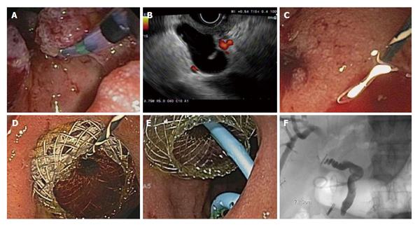
Figure 2 A 84 years old male with duodenal adenocarcinoma causing biliary obstruction underwent endoscopic ultrasound-guided choledochoduodenostomy following unsuccessful endoscopic retrograde cholangiopancreatography.
A: Tumor involving the major papilla; B: Endoscopic ultrasound-guided puncture of the common bile duct through the duodenum with a 19-gauge needle; C: Guidewire insertion and balloon dilation of a choledochoduodenal fistula; D: Followed by the placement of a 10 mm × 10 mm lumen-apposing metal stent to create a choledochoduodenostomy; E and F: Endoscopic (E) and flouroscopic (F) view after the placement of a 7 Fr × 3 cm double pigtail stent into the common hepatic duct.
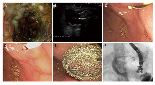
Figure 3 A 66 years old female with metastatic cholangiocarcinoma and gastric outlet obstruction undergoing endoscopic ultrasound-guided gastrojejunostomy.
A: Tumor ingrowth into two previously placed duodenal stents; B: Endoscopic ultrasound visualization of a 20 mm balloon inflated in the proximal jejunum followed by a 19-gauge needle puncture (arrow); C and D: Balloon dilation of the gastrojejunal fistula over a 0.035 inch guidewire; E and F: Endoscopic (E) and fluoroscopic (F) demonstration of contrast flow across 10 mm × 15 mm lumen-apposing metal stent (arrow) into the jejunum.
Figure 4 Magnetic anastomosis device to create endoscopic gastrojejunostomy (Images courtesy of Cook Medical).
A: Gastric magnent marked with an endoscopy clip; B: Mating of gastric and proximal jejunal magnets under fluoroscopic guidance to create a gastrojejunal fistula; C: Placement of a fully covered stent within the fistula with a proximal flanged edge positioned in the gastric lumen; D: The stent within the fistula functions as a gastrojejunostomy.
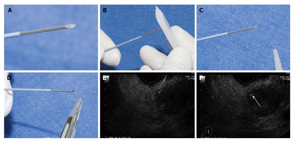
Figure 5 Images courtesy of Sanders et al[74].
A: Fiducial loaded into 19-gauge needle with sterile forceps; B: Fiducial within tip of needle; C: Sealing fiducial with sterile bonewax; D: Loaded fiducial ready for advancement down operating channel; E and F: Needle delivering fiducial into pancreatic mass (arrow).
- Citation: Oh SY, Irani S, Kozarek RA. What are the current and potential future roles for endoscopic ultrasound in the treatment of pancreatic cancer? World J Gastrointest Endosc 2016; 8(7): 319-329
- URL: https://www.wjgnet.com/1948-5190/full/v8/i7/319.htm
- DOI: https://dx.doi.org/10.4253/wjge.v8.i7.319


