Copyright
©The Author(s) 2016.
World J Gastrointest Endosc. Jan 25, 2016; 8(2): 30-39
Published online Jan 25, 2016. doi: 10.4253/wjge.v8.i2.30
Published online Jan 25, 2016. doi: 10.4253/wjge.v8.i2.30
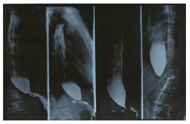
Figure 1 Barium swallow esophagrams showing typical bird-beak appearance of the distal esophagus.
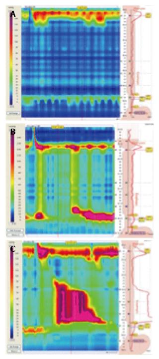
Figure 2 High-resolution manometric types of achalasia according to the Chicago Classification: Type 1, characterized by the absence of peristalsis (A); type 2, defined by the presence of esophageal compression, named panesophageal pressurization (B); type 3, characterized by the presence of peristaltic fragments or spastic waves (C).
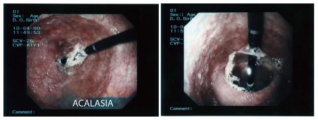
Figure 3 Pneumatic dilation with a Rigiflex balloon under endoscopic control.
- Citation: Esposito D, Maione F, D’Alessandro A, Sarnelli G, De Palma GD. Endoscopic treatment of esophageal achalasia. World J Gastrointest Endosc 2016; 8(2): 30-39
- URL: https://www.wjgnet.com/1948-5190/full/v8/i2/30.htm
- DOI: https://dx.doi.org/10.4253/wjge.v8.i2.30


