Copyright
©The Author(s) 2015.
World J Gastrointest Endosc. Jun 10, 2015; 7(6): 659-664
Published online Jun 10, 2015. doi: 10.4253/wjge.v7.i6.659
Published online Jun 10, 2015. doi: 10.4253/wjge.v7.i6.659
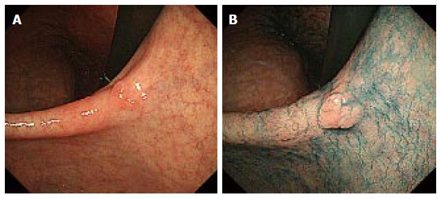
Figure 1 A case of a delineating lesion (0-IIa).
Before (A) and after (B) indigo-carmine dye, the margin of the tumor was clear.
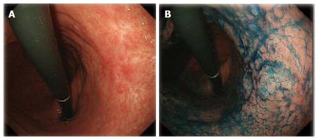
Figure 2 An undelineated margin lesion.
A: A case of an undelineating lesion (0-IIc) with ulceration findings; B: After indigo-carmine dye, the margin of the tumor was still unclear.
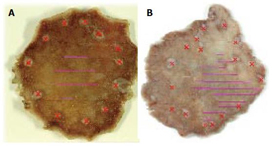
Figure 3 Cases of accurate and inaccurate markings.
A: A case of accurate markings. The purple lines indicate the tumor area. The red crosses indicate the marking; B: A case of inaccurate markings. The purple lines indicate the tumor area. The red crosses indicate the marking.
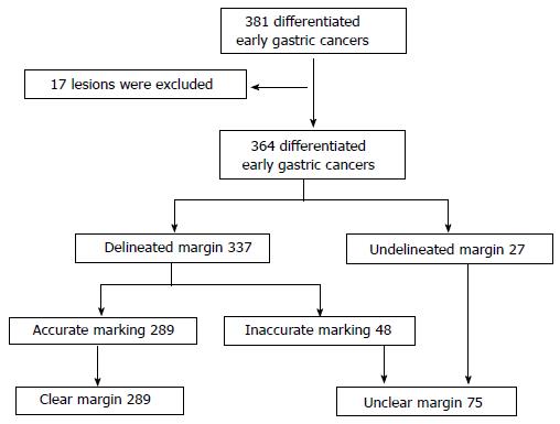
Figure 4 Flow chart of this study.
- Citation: Yoshinaga S, Oda I, Abe S, Nonaka S, Suzuki H, Takisawa H, Taniguchi H, Saito Y. Evaluation of the margins of differentiated early gastric cancer by using conventional endoscopy. World J Gastrointest Endosc 2015; 7(6): 659-664
- URL: https://www.wjgnet.com/1948-5190/full/v7/i6/659.htm
- DOI: https://dx.doi.org/10.4253/wjge.v7.i6.659


