©The Author(s) 2015.
World J Gastrointest Endosc. Apr 16, 2015; 7(4): 336-345
Published online Apr 16, 2015. doi: 10.4253/wjge.v7.i4.336
Published online Apr 16, 2015. doi: 10.4253/wjge.v7.i4.336
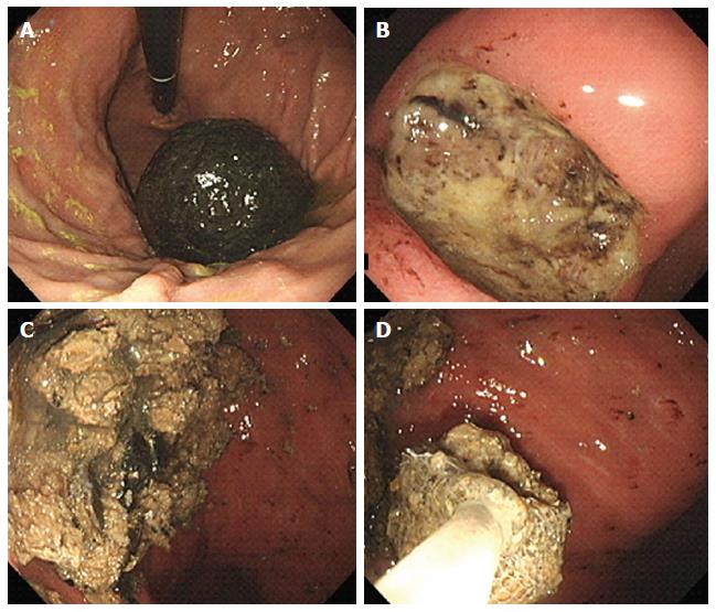
Figure 1 Endoscopic images of a persimmon phytobezoar.
A: A large, black bezoar is seen in the gastric fundus; B: A peptic ulcer is also observed in the gastric angle; C: Fragmentation of the bezoar was performed by endoscopy forceps and polypectomy snares; D: The fragments were removed by a retrieval net device and used in the subsequent in vitro analysis.
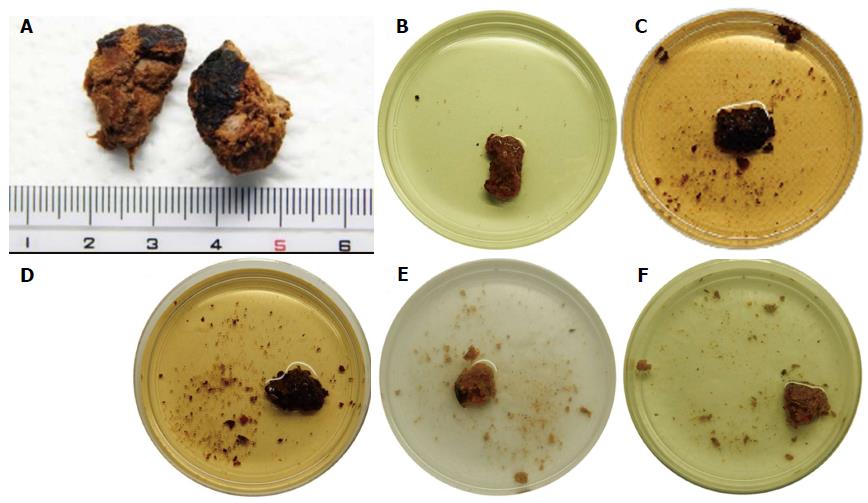
Figure 2 Photographs of the in vitro experiment.
A: Endoscopically extracted fragments of the gastric bezoar were used; B: Representative photographs of the bezoar fragments incubated at 37 °C with gentle swirling for 12 h with double-distilled water; C: Bezoar fragments after incubation with Coca-Cola®; D: Bezoar fragments after incubation with Coca-Cola Zero®; E: Bezoar fragments after incubation with a digestive enzymes supplement including cellulase; F: Bezoar fragments after incubation with papain. The bezoar fragments were clearly more softened and more fragmented after 12-h incubation with Coca-Cola® or Coca-Cola Zero® than with the other agents.
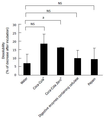
Figure 3 The dissolubility of bezoar fragments.
The mean dissolubility of bezoar fragments was highest by Coca-Cola®, but the difference between Coca-Cola® and water was not significant (P = 0.06) due to the relatively large standard deviation. NS: Not significant. aP < 0.05.
- Citation: Iwamuro M, Okada H, Matsueda K, Inaba T, Kusumoto C, Imagawa A, Yamamoto K. Review of the diagnosis and management of gastrointestinal bezoars. World J Gastrointest Endosc 2015; 7(4): 336-345
- URL: https://www.wjgnet.com/1948-5190/full/v7/i4/336.htm
- DOI: https://dx.doi.org/10.4253/wjge.v7.i4.336


