©The Author(s) 2015.
World J Gastrointest Endosc. Sep 10, 2015; 7(12): 1055-1061
Published online Sep 10, 2015. doi: 10.4253/wjge.v7.i12.1055
Published online Sep 10, 2015. doi: 10.4253/wjge.v7.i12.1055
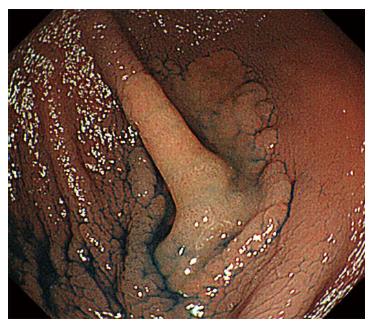
Figure 1 Chromoendoscopy showing a 30 mm laterally spreading tumor (non-granular type) located at the bottom of the cecum bottom.
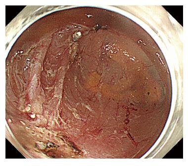
Figure 2 Endoscopic submucosal dissection is performed safely without intraoperative perforation.
The procedure time is 47 min.
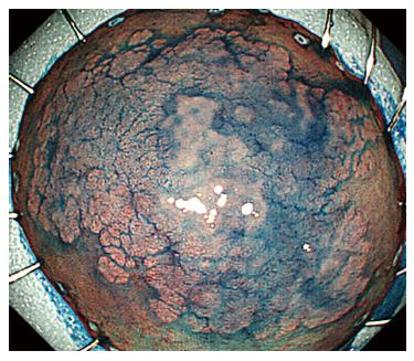
Figure 3 The tumor is resected in an en bloc fashion, and the specimen measures 51 mm × 40 mm.
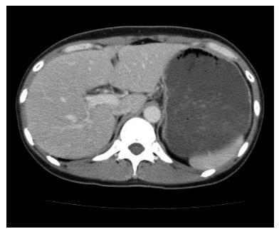
Figure 4 Abdominal contrast enhanced computed tomography 24 h after endoscopic submucosal dissection.
Free air is not recognized.
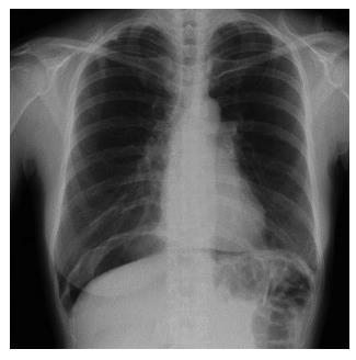
Figure 5 Abdominal radiography 36 h after endoscopic submucosal dissection because of a complaint of severe abdominal pain.
A large amount of free air is observed under the diaphragm.
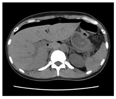
Figure 6 Abdominal computed tomography 36 h after endoscopic submucosal dissection.
A large amount of free air is observed on the surface of the liver and the intraperitoneal cavity.
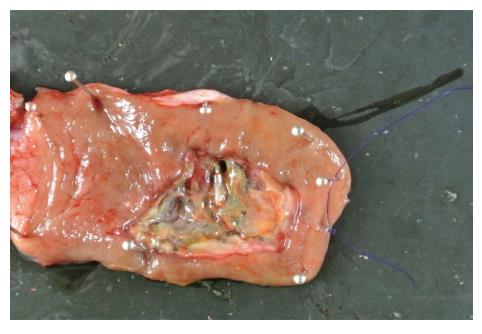
Figure 7 A small perforation site is present in the surgical specimen.
- Citation: Hirasawa K, Sato C, Makazu M, Kaneko H, Kobayashi R, Kokawa A, Maeda S. Coagulation syndrome: Delayed perforation after colorectal endoscopic treatments. World J Gastrointest Endosc 2015; 7(12): 1055-1061
- URL: https://www.wjgnet.com/1948-5190/full/v7/i12/1055.htm
- DOI: https://dx.doi.org/10.4253/wjge.v7.i12.1055


