Copyright
©2014 Baishideng Publishing Group Inc.
World J Gastrointest Endosc. Aug 16, 2014; 6(8): 345-351
Published online Aug 16, 2014. doi: 10.4253/wjge.v6.i8.345
Published online Aug 16, 2014. doi: 10.4253/wjge.v6.i8.345
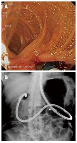
Figure 1 View Billroth II.
A: Endoscopic view of Billroth II gastrojejunostomy with sharp angulation towards the afferent limb while retracting the single-balloon enteroscope. Note the mucosal tear at the short angle of the afferent limb at the end of the endoscopic retrograde cholangiopancreatography procedure; B: Radiological view of the looping position of the single-balloon enteroscope in the stomach of a patient with Billroth II partial gastrectomy. The tip of the enteroscope is located in the blind end of the duodenum. The white arrow denotes the position of the deflated overtube balloon.
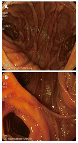
Figure 2 Endoscopic view of Roux-en-Y reconstruction.
A: Endoscopic view of an end-to-side Roux-en-Y reconstruction with view on the afferent limb (right) and common limb (left). The white arrow denotes the operative scar of the anastomosis; B: Endoscopic view of a side-to-side Roux-en-Y reconstruction with view on the afferent limb (middle), common limb (left) and blind ending limb (right). The white arrow denotes the operative scar of the anastomosis.
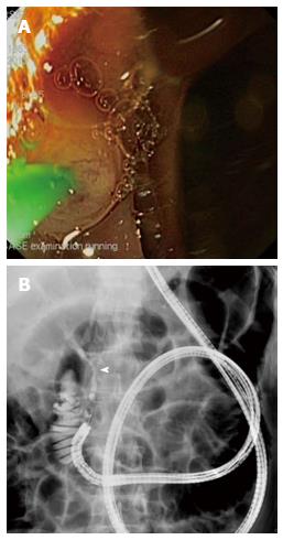
Figure 3 View.
A: Endoscopic view of the distal approach to cannulate an intact papilla of Vater using a straight catheter with a forward-viewing endoscope. In order to cannulate in the direction of the common bile duct, the papilla is rotated into the 7 o’clock position; B: Radiological view of the double-balloon enteroscope in a patient with Roux-en-Y gastric bypass. The distal approach with a forward-viewing endoscope allows straight cannulation of the common bile duct (white arrow) in line with the direction of the working channel.
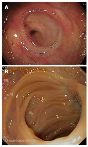
Figure 4 Endoscopic view of bilioenteric anastomosis.
A: Endoscopic view of a normal end-to-side bilioenteric anastomosis; B: Endoscopic view of a stenosis at the level of the bilioenteric anastomosis. Only scar tissue (white arrow) indicates the location of the anastomosis without visible opening.
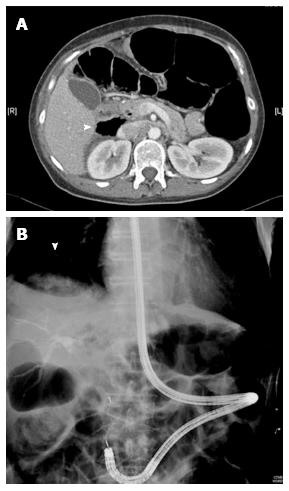
Figure 5 A patient with Roux-en-Y gastric bypass.
A: Computed tomography of a retroperitoneal perforation (white arrow) at the level of papilla of Vater after sphincterotomy and sphincteroplasty in a patient with Roux-en-Y gastric bypass; B: Radiological view of hepatic capsule dehiscence without free abdominal air (white arrow) due to barotrauma in the closed afferent limb during single-balloon enteroscopy endoscopic retrograde cholangiopancreatography in a patient with Billroth II partial gastrectomy. Common bile duct stone retrieval with a basket is being performed.
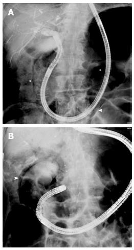
Figure 6 Radiological view.
A: Radiological view of direct cholangioscopy with the single-balloon enteroscope inside the common bile duct and with the overtube (white arrow) inside the afferent limb of a Billroth II patient; B: Radiological view of free air around the contrast-filled gallbladder (white arrow) due to barotrauma during single-balloon enteroscopy endoscopic retrograde cholangiopancreatography in a Billroth II patient.
- Citation: Moreels TG. Endoscopic retrograde cholangiopancreatography in patients with altered anatomy: How to deal with the challenges? World J Gastrointest Endosc 2014; 6(8): 345-351
- URL: https://www.wjgnet.com/1948-5190/full/v6/i8/345.htm
- DOI: https://dx.doi.org/10.4253/wjge.v6.i8.345


