Copyright
©2013 Baishideng Publishing Group Co.
World J Gastrointest Endosc. Sep 16, 2013; 5(9): 420-427
Published online Sep 16, 2013. doi: 10.4253/wjge.v5.i9.420
Published online Sep 16, 2013. doi: 10.4253/wjge.v5.i9.420
Figure 1 Per-oral endoscopic myotomy technique (© Winthrop University Hospital, 2012).
A mucosal incision is performed after submucosal injection. Then, dissection of the submucosal tunnel is initiated and extended into the gastric cardia. Myotomy is performed of the circular layer and extended 2 cm into the gastric cardia. Finally, the entrance to the submucosal tunnel is closed with endoclips.
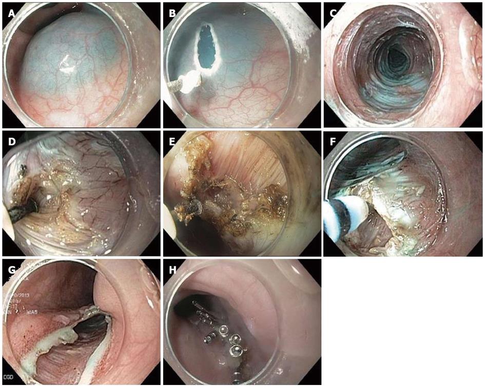
Figure 2 Per-oral endoscopic myotomy technique.
A: Submucosal injection; B: Mucosal incision; C: Submucosal tunnel; D: Myotomy initiation; E: Dissection of the circular layer with longitudinal layer intact; F: Hooking of circular muscle layer with hybrid knife; G: Entrance to the submucosal tunnel; H: Closure.
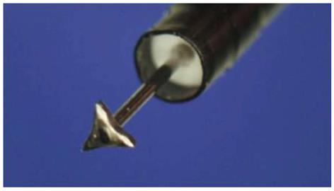
Figure 3 Triangular tip knife (Olympus) Courtesy Haruhiro Inoue.
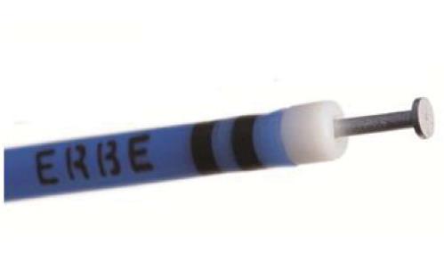
Figure 4 T-type hybrid knife (ERBE Elektromedizin GmbH).
Courtesy John Day, ERBE (Marietta, GA).
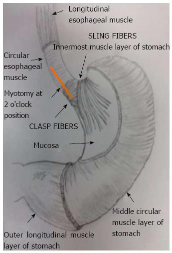
Figure 5 Human lower esophageal sphincter anatomy in relation to per-oral endoscopic myotomy myotomy orientation (© S.
N. Stavropoulos, Winthrop University Hospital, 2012).
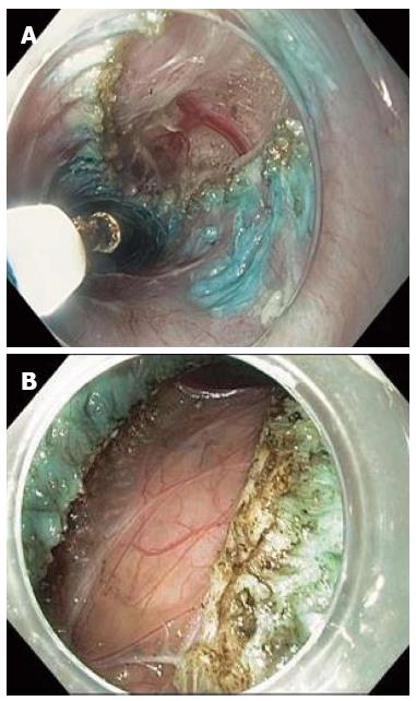
Figure 6 Full-thickness myotomy.
- Citation: Friedel D, Modayil R, Iqbal S, Grendell JH, Stavropoulos SN. Per-oral endoscopic myotomy for achalasia: An American perspective. World J Gastrointest Endosc 2013; 5(9): 420-427
- URL: https://www.wjgnet.com/1948-5190/full/v5/i9/420.htm
- DOI: https://dx.doi.org/10.4253/wjge.v5.i9.420


