Copyright
©2013 Baishideng Publishing Group Co.
World J Gastrointest Endosc. Apr 16, 2013; 5(4): 141-147
Published online Apr 16, 2013. doi: 10.4253/wjge.v5.i4.141
Published online Apr 16, 2013. doi: 10.4253/wjge.v5.i4.141
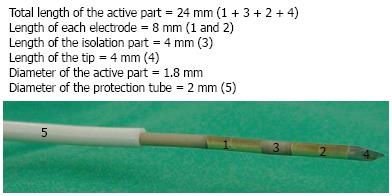
Figure 1 The tip of the ERBE hybrid cryotherm probe with the active electrical part.
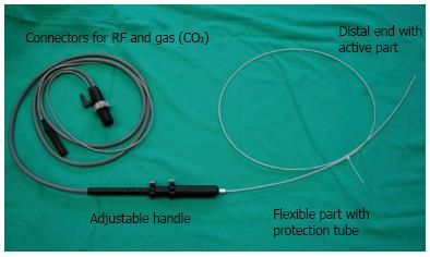
Figure 2 The ERBE flexible probe used for endoscopic ultrasound-guided ablation of the parenchymal organs.
The probe, covered with a protection tube, is passed through the operative channel of the echoendoscope.
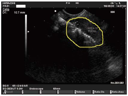
Figure 3 The cryotherm probe applied in the porcine pancreas: the probe is seen as an hyperechoic line.
Initially an hyperechoic elliptic area appears around the distal tip of the probe, surrounded by a hypoechoic border (most likely edema).
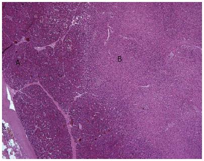
Figure 4 Histopathologic section from the first pig: Normal pancreatic tissue (A) surrounds the central treated area (B).
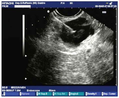
Figure 5 Endoscopic ultrasound-guided puncture of a cystic tumor.
The cyst is punctured with a 22-G fine needle under endoscopic ultrasound guidance, the fluid is aspirated, then ethanol is injected into the cyst and re-aspirated after 3-5 min.
- Citation: Carrara S, Petrone MC, Testoni PA, Arcidiacono PG. Tumors and new endoscopic ultrasound-guided therapies. World J Gastrointest Endosc 2013; 5(4): 141-147
- URL: https://www.wjgnet.com/1948-5190/full/v5/i4/141.htm
- DOI: https://dx.doi.org/10.4253/wjge.v5.i4.141


