Copyright
©2013 Baishideng Publishing Group Co.
World J Gastrointest Endosc. Oct 16, 2013; 5(10): 487-494
Published online Oct 16, 2013. doi: 10.4253/wjge.v5.i10.487
Published online Oct 16, 2013. doi: 10.4253/wjge.v5.i10.487
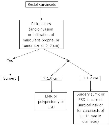
Figure 1 Treatment of rectal carcinoids.
EMR: Endoscopic mucosal resection; ESD: Endoscopic submucosal dissection.
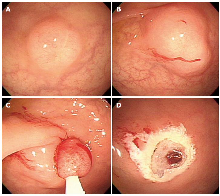
Figure 2 Endoscopic mucosal resection.
A: An approximately 6 mm rectal carcinoid tumor; B: Injection of submucosal saline solution; C: Endoscopic mucosal resection (EMR) procedure; D: A clear, post-EMR ulcer.
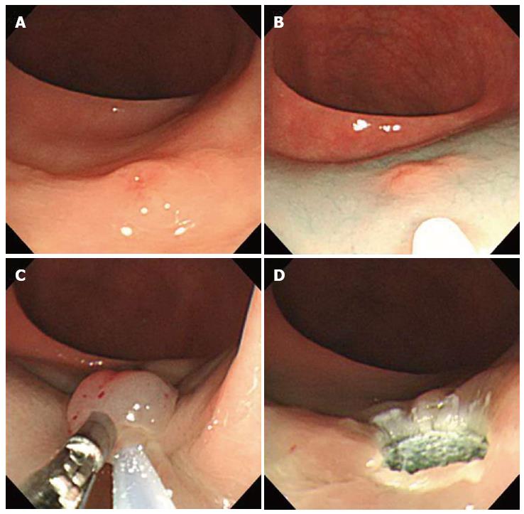
Figure 3 Endoscopic mucosal resection using two-channel colonoscopy.
A: An approximately 5 mm rectal carcinoid tumor; B: Injection of submucosal saline solution into the base of the lesion using needle forcep; C: Pulling the lesion with grasping forcep and snare resection; D: A clear, post-endoscopic mucosal resection ulcer.
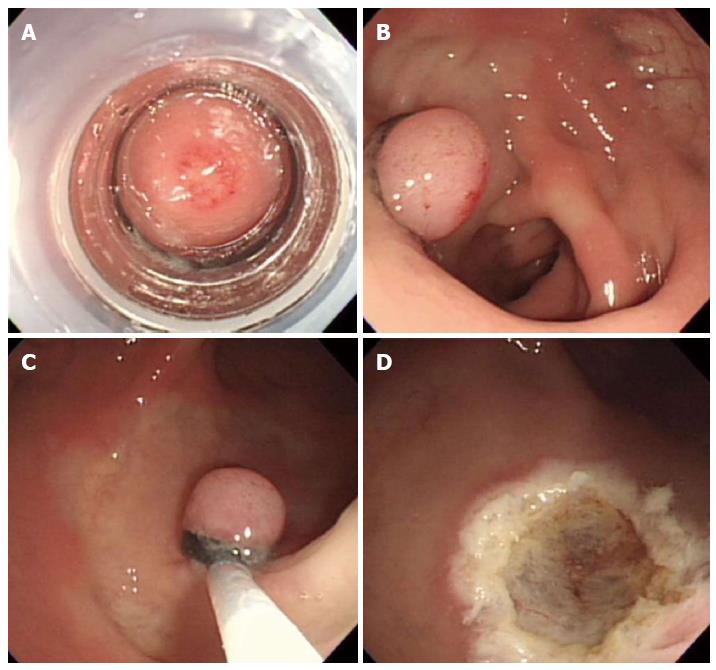
Figure 4 Endoscopic submucosal resection with ligating device.
A: Aspiration of a carcinoid tumor into the ligating device; B: Deployed elastic band; C: Snare resection performed below the band; D: A clear, post-endoscopic submucosal resection with ligating device ulcer.
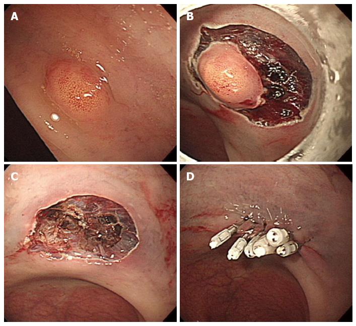
Figure 5 Endoscopic submucosal dissection.
A: An approximately 5 mm rectal carcinoid tumor; B: Mucosal incision and submucosal dissection; C: A clear, post-endoscopic submucosal dissection ulcer; D: Endoscopic closure of the ulcer floor with endoclips.
- Citation: Choi HH, Kim JS, Cheung DY, Cho YS. Which endoscopic treatment is the best for small rectal carcinoid tumors? World J Gastrointest Endosc 2013; 5(10): 487-494
- URL: https://www.wjgnet.com/1948-5190/full/v5/i10/487.htm
- DOI: https://dx.doi.org/10.4253/wjge.v5.i10.487


