Copyright
©20???? Baishideng Publishing Group Co.
World J Gastrointest Endosc. Dec 16, 2012; 4(12): 571-574
Published online Dec 16, 2012. doi: 10.4253/wjge.v4.i12.571
Published online Dec 16, 2012. doi: 10.4253/wjge.v4.i12.571
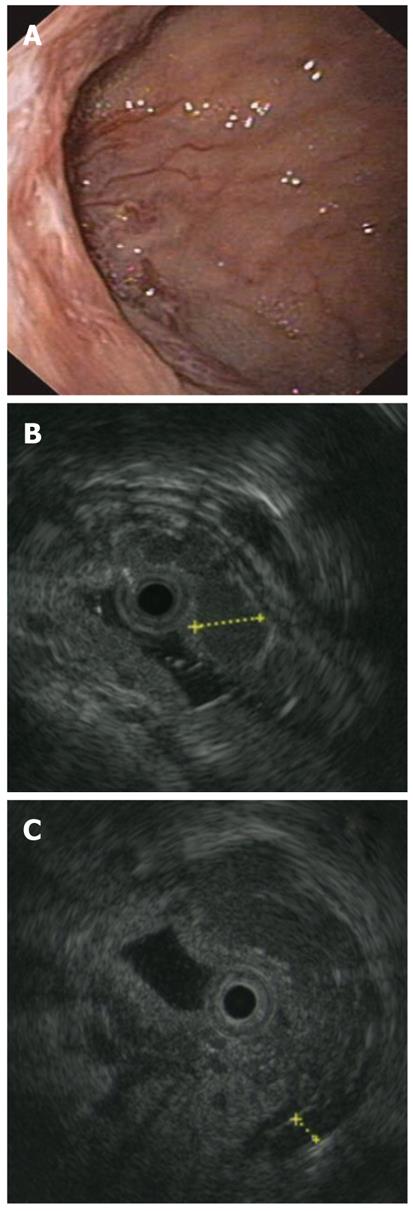
Figure 1 Lesions in the duodenum.
A: Depressed area of 2 cm, covered with mixed hyperemic and white mucosa, seen at EGDS; B: Round hypo-anechoic areas of 5 mm in diameter, compatible with vascular structures (varices), in the duodenal mucosa; C: Periduodenal anechoic lesions compatible with collaterals.
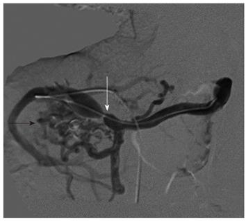
Figure 2 Transhepatic portography showing a severe anastomotic stenosis (arrow), and filling of multiple periduodenal varices (arrow head).
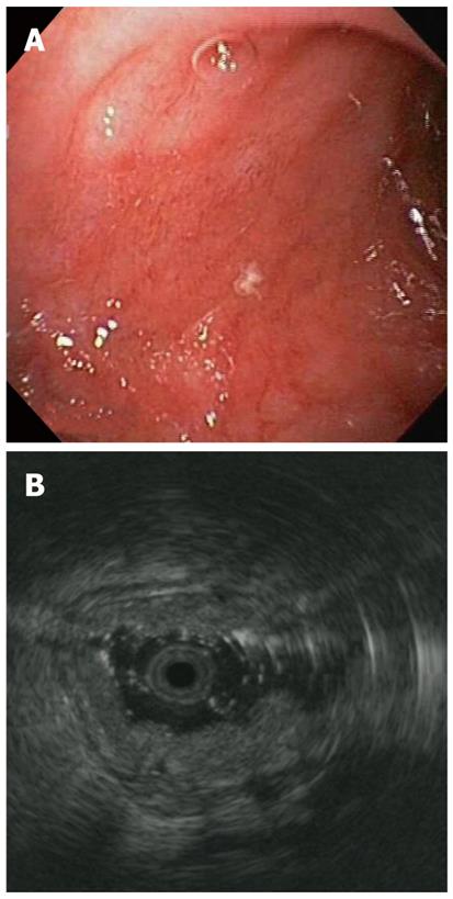
Figure 3 Normal endoscopic appearance of the bulb (A) and normal stratification of the duodenal layers (B).
- Citation: Curcio G, Pisa MD, Miraglia R, Catalano P, Barresi L, Tarantino I, Granata A, Spada M, Traina M. Case of obscure-overt gastrointestinal bleeding after pediatric liver transplantation explained by endoscopic ultrasound. World J Gastrointest Endosc 2012; 4(12): 571-574
- URL: https://www.wjgnet.com/1948-5190/full/v4/i12/571.htm
- DOI: https://dx.doi.org/10.4253/wjge.v4.i12.571


