©2012 Baishideng Publishing Group Co.
World J Gastrointest Endosc. Jan 16, 2012; 4(1): 9-16
Published online Jan 16, 2012. doi: 10.4253/wjge.v4.i1.9
Published online Jan 16, 2012. doi: 10.4253/wjge.v4.i1.9
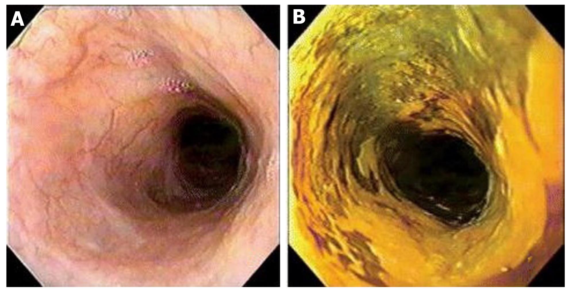
Figure 1 Conventional esophagoscopy and Lugol’s chromoendoscopy.
A: Conventional esophagoscopy presenting normal appearing mucosa; B: Lugol’s chromoendoscopy disclosed an unstained areaafter multiple biopsies, the diagnosis was high-grade dysplasia.
- Citation: Lopes AB, Fagundes RB. Esophageal squamous cell carcinoma - precursor lesions and early diagnosis. World J Gastrointest Endosc 2012; 4(1): 9-16
- URL: https://www.wjgnet.com/1948-5190/full/v4/i1/9.htm
- DOI: https://dx.doi.org/10.4253/wjge.v4.i1.9


