©2012 Baishideng Publishing Group Co.
World J Gastrointest Endosc. Jan 16, 2012; 4(1): 1-8
Published online Jan 16, 2012. doi: 10.4253/wjge.v4.i1.1
Published online Jan 16, 2012. doi: 10.4253/wjge.v4.i1.1
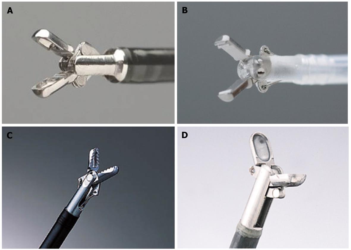
Figure 1 Hemostatic forceps tips.
A: Monopolar hemostatic forceps (HDB2422W; Pentax, Tokyo, Japan); B: Bipolar hemostaticforceps (H-S2518; Pentax, Tokyo, Japan); C: Hemostatic forceps (Coagrasper: FD-410LR; Olympus, Tokyo, Japan); D: Hot biopsy forceps (FD-1L-1; Olympus, Tokyo, Japan).
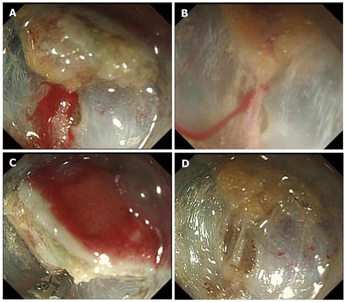
Figure 2 Hemostatic procedure for endoscopic submucosal dissectionintraoperative bleeding using hemostatic forceps.
A: Pulsatile bleeding is observed during submucosal dissection; B: By filling the tip attachment with water, the bleeding point can be pinpointed and identified; C: After identifying the bleeding point, the vessel is securely grasped by hemostatic forceps, and thermo-coagulation is performed; D: Complete hemostasis is achieved, without excessive coagulation.
- Citation: Muraki Y, Enomoto S, Iguchi M, Fujishiro M, Yahagi N, Ichinose M. Management of bleeding and artificial gastric ulcers associated with endoscopic submucosal dissection. World J Gastrointest Endosc 2012; 4(1): 1-8
- URL: https://www.wjgnet.com/1948-5190/full/v4/i1/1.htm
- DOI: https://dx.doi.org/10.4253/wjge.v4.i1.1


