Copyright
©2011 Baishideng Publishing Group Co.
World J Gastrointest Endosc. Nov 16, 2011; 3(11): 235-240
Published online Nov 16, 2011. doi: 10.4253/wjge.v3.i11.235
Published online Nov 16, 2011. doi: 10.4253/wjge.v3.i11.235
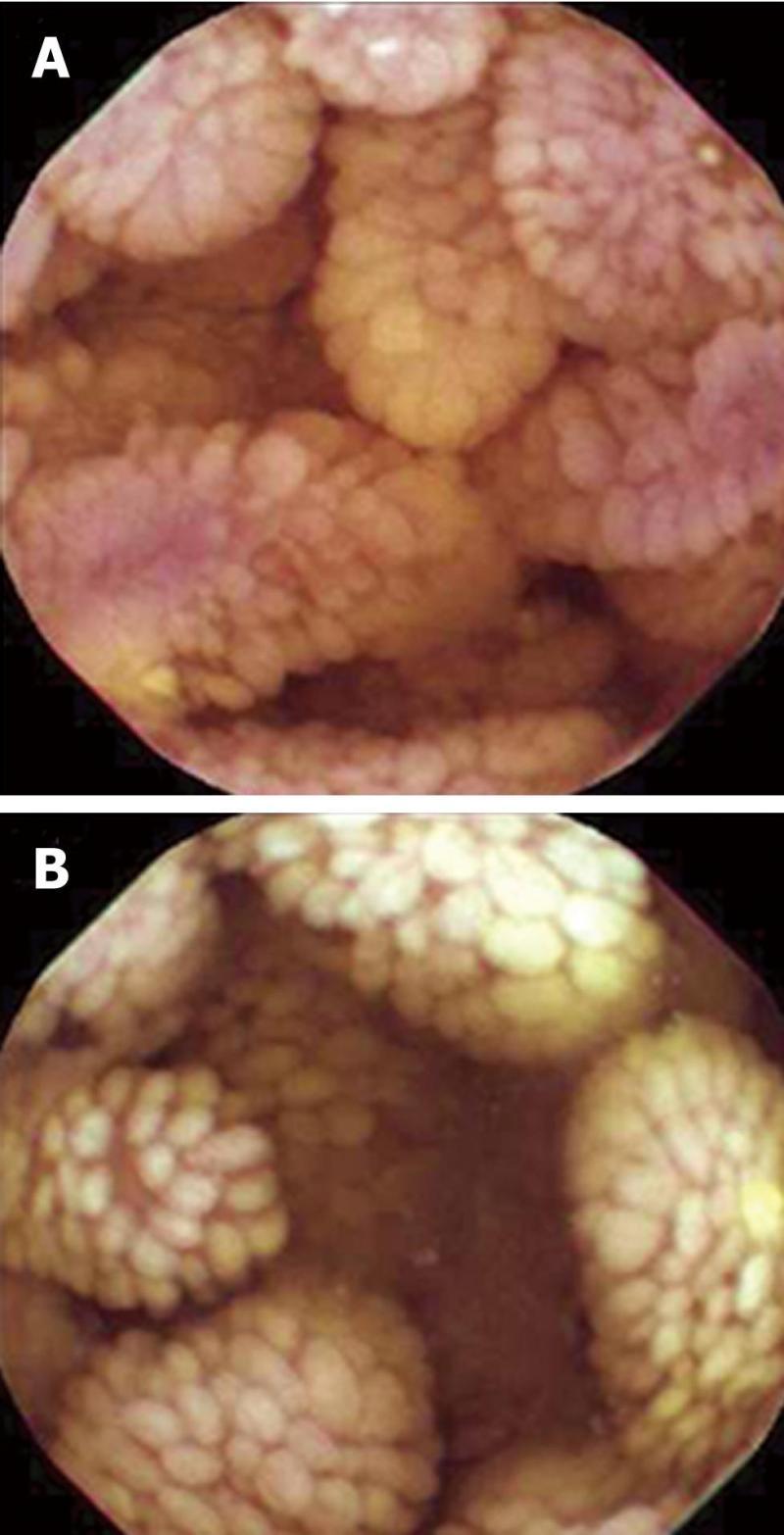
Figure 1 Capsule endoscopy shows diffuse edematous mucosae covered with enlarged and swollen villi in the jejunum (A), and diffuse finger-like elongated mucosa covered with enlarged whitish villi in the ileum (B).
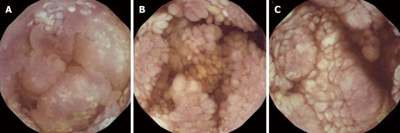
Figure 2 Capsule endoscopic findings in the duodenum (A), jejunum (B) and ileum (C).
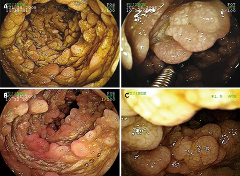
Figure 3 Double balloon enteroscopy was performed with biopsy forceps to obtain a small bowel specimen of polypoid mucosa covered with enlarged whitish villi (A); and additional double balloon enteroscopic findings (B, C).
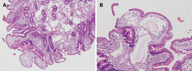
Figure 4 Histology of mucosal tissue in the jejunum shows multiple dilated lymphatics.
A: HE, × 40; B: HE, × 100.
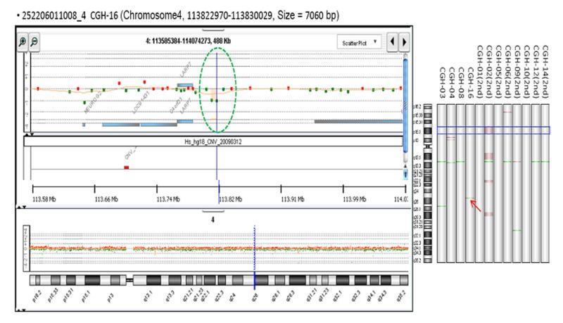
Figure 5 The deletion on chromosome 4q25.
- Citation: Oh TG, Chung JW, Kim HM, Han SJ, Lee JS, Park JY, Song SY. Primary intestinal lymphangiectasia diagnosed by capsule endoscopy and double balloon enteroscopy. World J Gastrointest Endosc 2011; 3(11): 235-240
- URL: https://www.wjgnet.com/1948-5190/full/v3/i11/235.htm
- DOI: https://dx.doi.org/10.4253/wjge.v3.i11.235


