Copyright
©2010 Baishideng.
World J Gastrointest Endosc. Aug 16, 2010; 2(8): 278-287
Published online Aug 16, 2010. doi: 10.4253/wjge.v2.i8.278
Published online Aug 16, 2010. doi: 10.4253/wjge.v2.i8.278
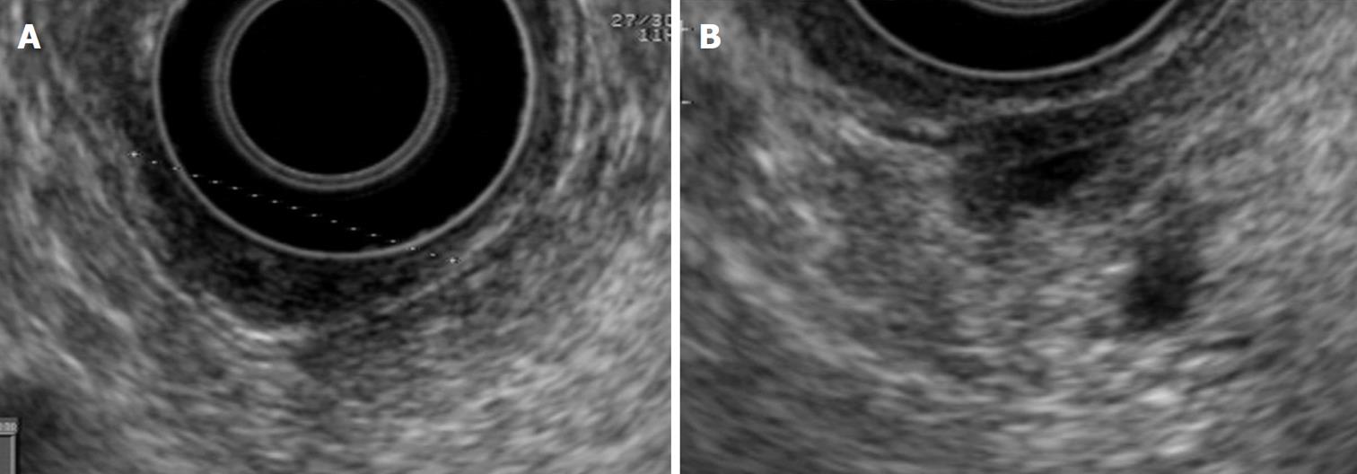
Figure 1 Normal radial view.
A: Ampulla; B: Periampullary region.
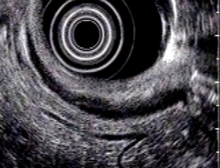
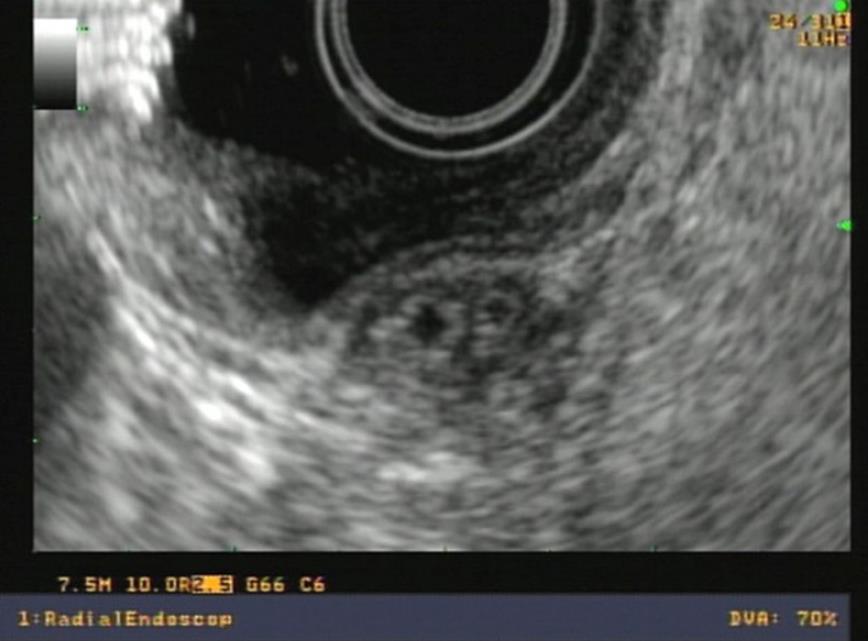
Figure 2 Radial view of normal distal choledochus and wirsung duct.
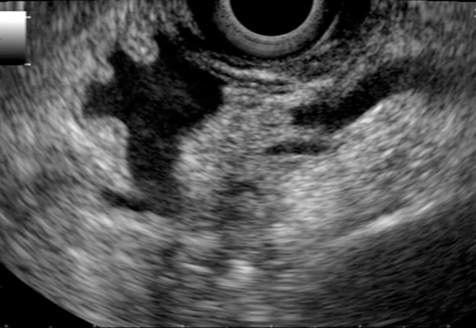
Figure 3 Linear view of normal papilla and periampullary region.
The opening of the choledochal and Wirsung ducts is seen through the papilla.
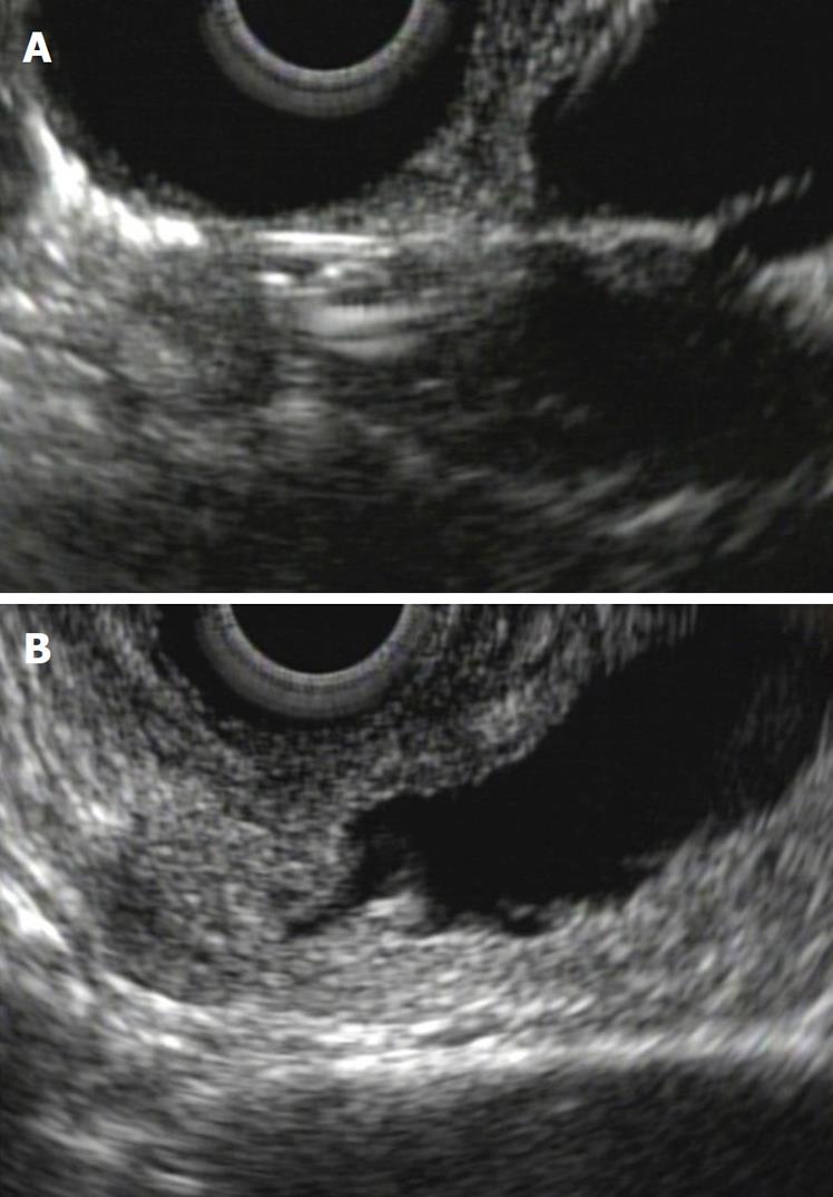
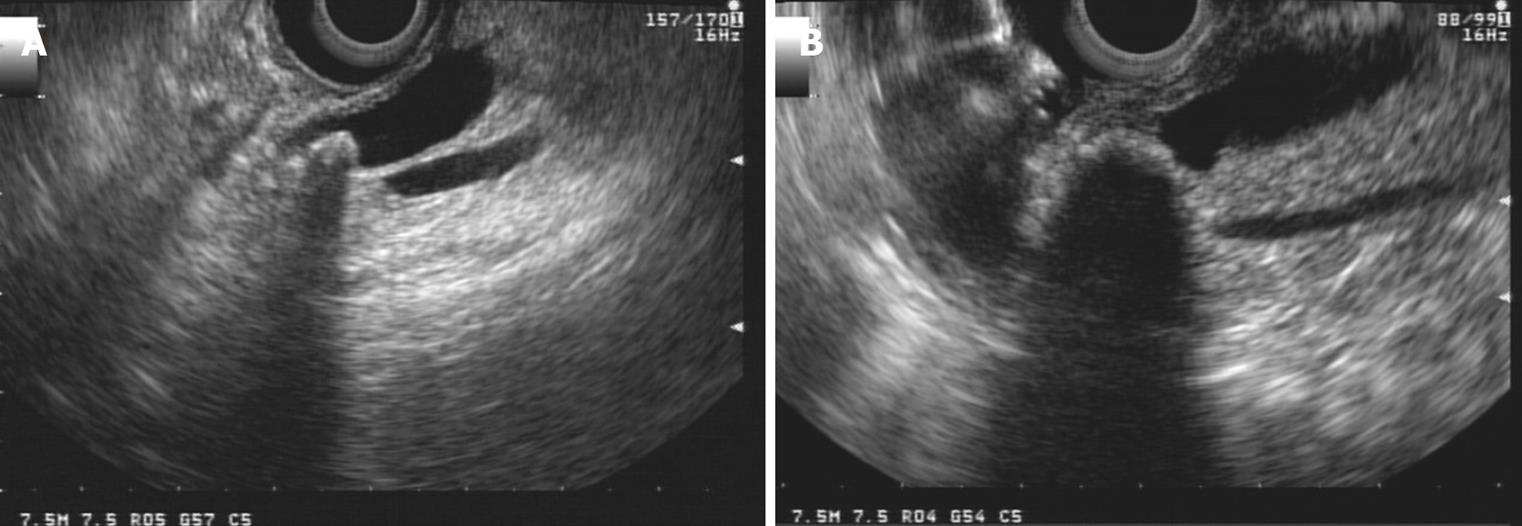
Figure 4 Biliary stent in the papilla, linear view.
A: Acoustic interference in periampullary region due to biliary stent; B: The same patient after stent removal.
Figure 5 Ampullary tubulo-villous adenoma.
A: Endoscopic view; B: Tumor restricted to the duodenal wall as shown in radial endoscopic ultrasound; C: No ductal ingrowth. Endoscopic ampullectomy was indicated.
Figure 6 Ampullary carcinoma.
A: Ulcerated ampullary tumor revealed by endoscopy; B: Duodenal wall disruption and pancreatic invasion in radial endoscopic ultrasound of the same tumor; C: Tumoral lymph nodes (same patient). Palliative therapy was indicated.
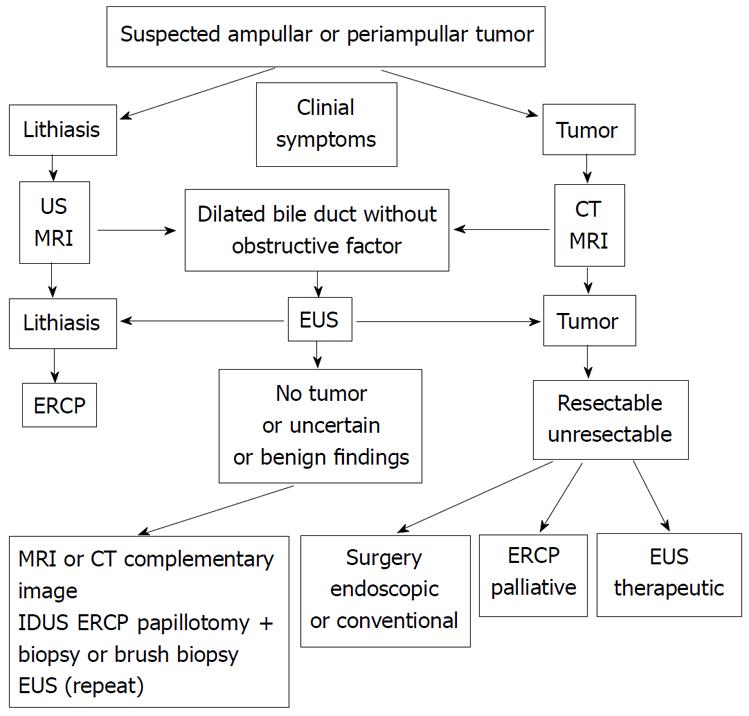
Figure 7 Procedural algorithm for suspected ampullary or periampullary tumor.
EUS: endoscopic ultrasound; MRI: magnetic resonance imaging; ERCP: endoscopic retrograde cholangiopancreatography; CT: computerized tomography; US: ultrasound; IDUS: intraductal sonography.
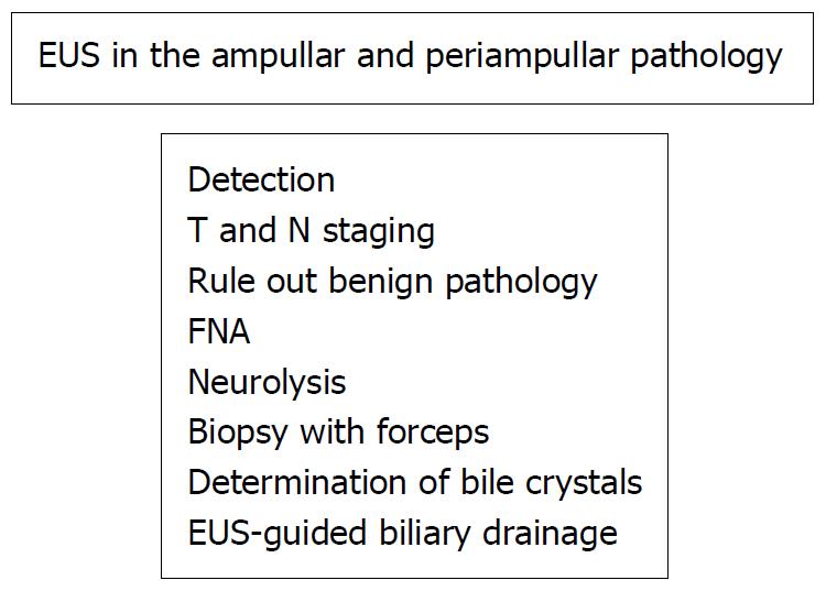
Figure 8 Role of the Endosonography in the ampullary and periampullary pathology.
EUS: endoscopic ultrasoun; FNA: fine needle aspiration.
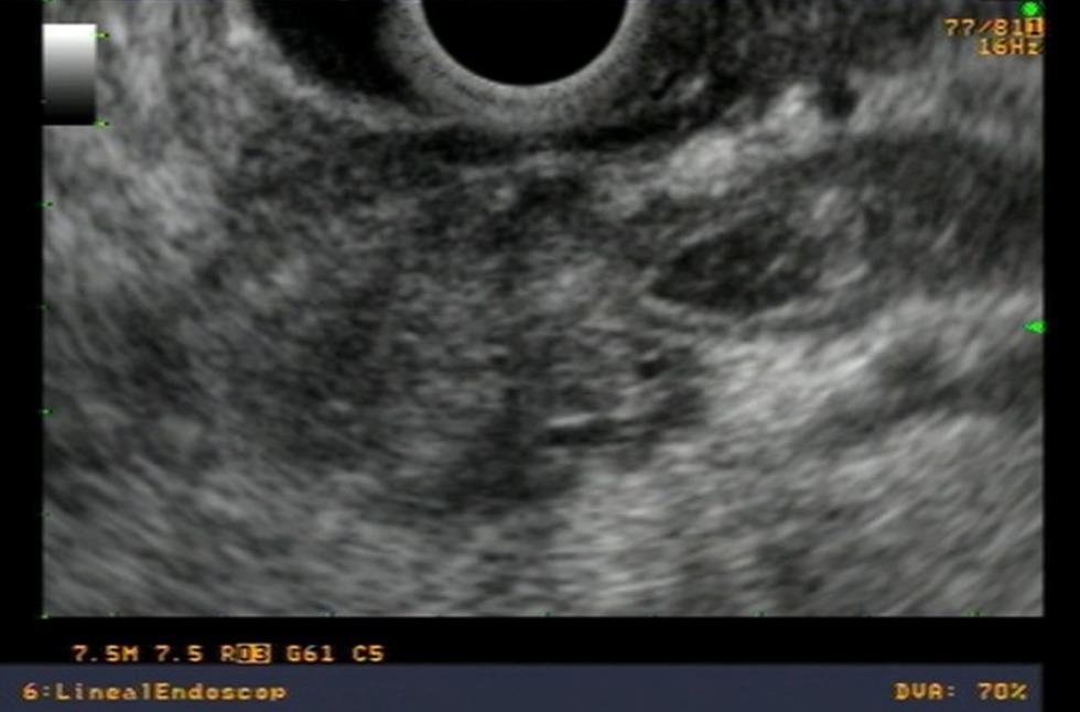
Figure 9 Linear view of periampullary carcinoma obstructing biliary duct.
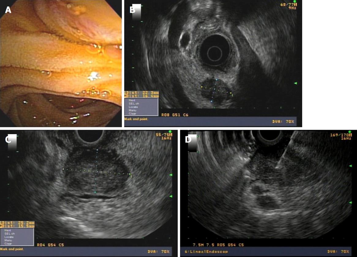
Figure 10 Periampullary tumor.
A: Normal papilla in endoscopic view; B: Radial endoscopic ultrasound (EUS) finding of periampullary tumor; C: Linear EUS view; D: Fine needle aspiration puncture.
Figure 11 Endoscopic ultrasound finding in acute pancreatitis in a patient with normal magnetic resonance imaging.
A: Impacted 4 mm stone in the ampulla with radial endoscopic ultrasound; B: Close-up; C: Precut during Endoscopic retrograde cholangiopancreatography in the same patient.
Figure 12 Lineal imaging of distal choledochal lithiasis.
A: Stone and acoustic shadow is clearly seen; B: Bulging of the papilla due to impacted stone.
Figure 13 Transverse view of the ampulla with radial transducer in a patient with suspected sphincter of oddi dysfunction.
Oddi muscle can be seen.
- Citation: Castillo C. Endoscopic ultrasound in the papilla and the periampullary region. World J Gastrointest Endosc 2010; 2(8): 278-287
- URL: https://www.wjgnet.com/1948-5190/full/v2/i8/278.htm
- DOI: https://dx.doi.org/10.4253/wjge.v2.i8.278


