©2010 Baishideng.
World J Gastrointest Endosc. Jul 16, 2010; 2(7): 268-270
Published online Jul 16, 2010. doi: 10.4253/wjge.v2.i7.268
Published online Jul 16, 2010. doi: 10.4253/wjge.v2.i7.268
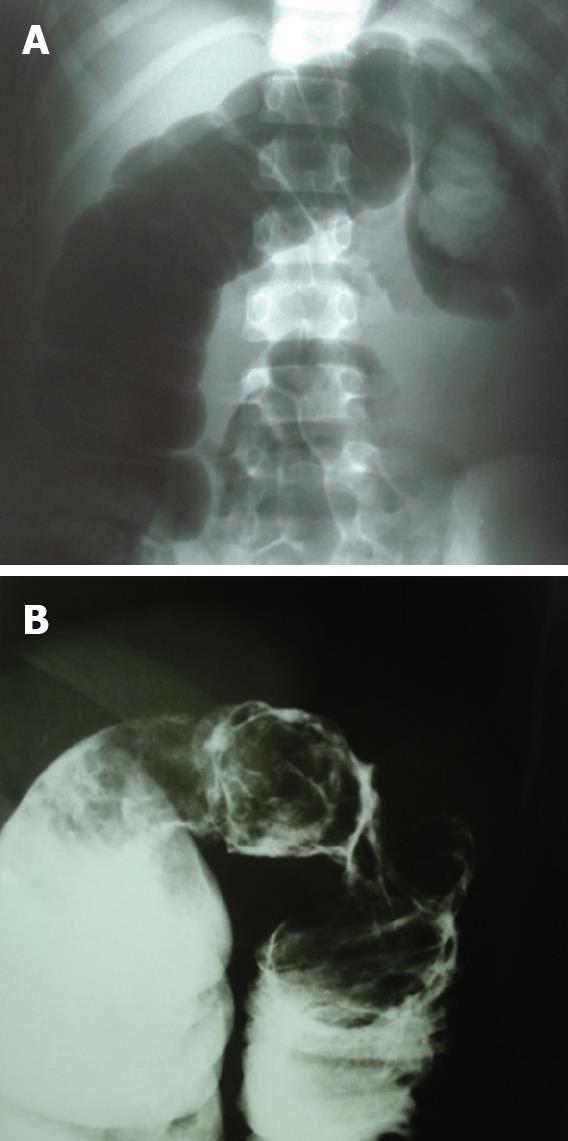
Figure 1 Plain abdominal film.
A: Completed colonic obstruction on the left side of the colon; B: Barium enema revealed a colonic mass at the splenic flexor after successful reduction.
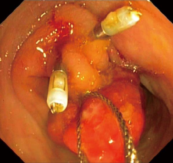
Figure 2 At the transverse colon, a huge colonic polyp with mucosal invagination confirmed that it was acting as a leading point.
Snare polypectomy was applied after successful vascular control with two hemoclips.
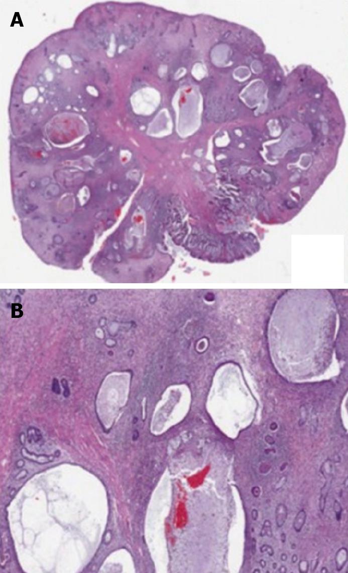
Figure 3 The whole mouth capture shows a polypoid mass, with edematous stroma, inflammation, and ulceration.
A: Multiple dilated mucous glands and mucin lakes are noted; B: The dilated mucous glands and stromal inflammation (× 2).
- Citation: Suksamanapun N, Uiprasertkul M, Ruangtrakool R, Akaraviputh T. Endoscopic treatment of a large colonic polyp as a cause of colocolonic intussusception in a child. World J Gastrointest Endosc 2010; 2(7): 268-270
- URL: https://www.wjgnet.com/1948-5190/full/v2/i7/268.htm
- DOI: https://dx.doi.org/10.4253/wjge.v2.i7.268


