©2010 Baishideng.
World J Gastrointest Endosc. Jun 16, 2010; 2(6): 203-209
Published online Jun 16, 2010. doi: 10.4253/wjge.v2.i6.203
Published online Jun 16, 2010. doi: 10.4253/wjge.v2.i6.203
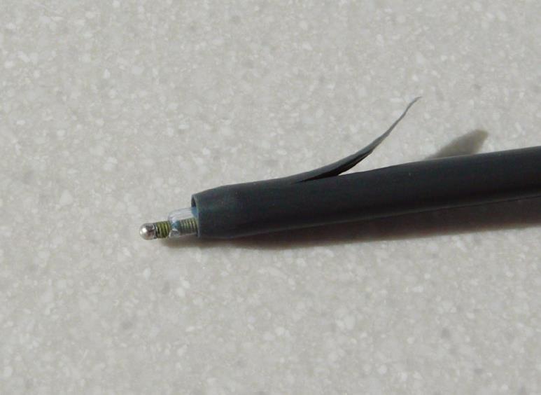
Figure 1 Photo of one-step device making the hole and placing one stent at the same time.
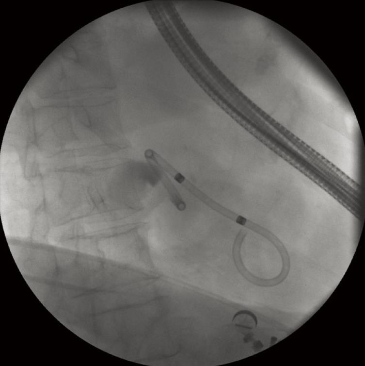
Figure 2 Double pigtail stent of 10 fr between the gallbladder and the gastric antrum.
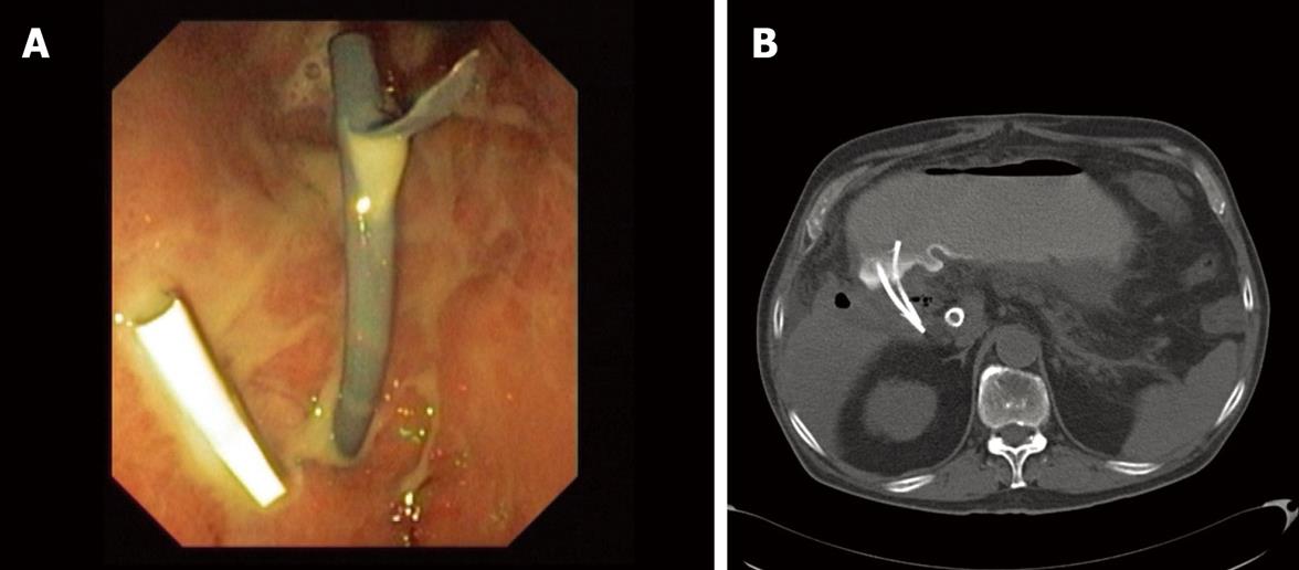
Figure 3 Two straight stents of 8.
5 fr draining purulent material between the gallbladder and the gastric antrum. A: From the gallbladder to the gastric antrum; B: CT-scan performed some days after the procedure showing the stents and bubbles of gas in the gallbladder fundus.
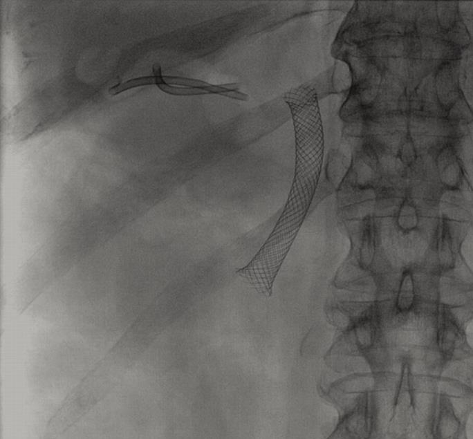
Figure 4 Detail of two straight stents of 8.
5 fr draining the gallbladder, and a metal stent in the common bile duct.
- Citation: Súbtil JC, Betes M, Muñoz-Navas M. Gallbladder drainage guided by endoscopic ultrasound. World J Gastrointest Endosc 2010; 2(6): 203-209
- URL: https://www.wjgnet.com/1948-5190/full/v2/i6/203.htm
- DOI: https://dx.doi.org/10.4253/wjge.v2.i6.203


