Copyright
©2010 Baishideng.
World J Gastrointest Endosc. May 16, 2010; 2(5): 179-185
Published online May 16, 2010. doi: 10.4253/wjge.v2.i5.179
Published online May 16, 2010. doi: 10.4253/wjge.v2.i5.179
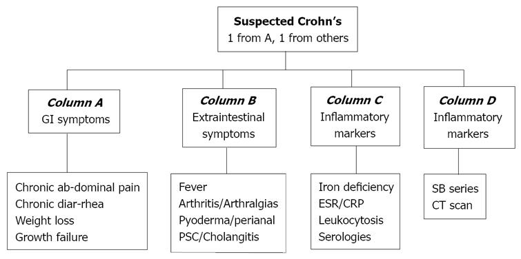
Figure 1 Criteria for suspected Crohn’s disease.
(Mergener et al[18]) (PSC: Primary sclerosing cholangitis; ESR: Erythrocyte sedimentation rate; CRP: C reactive protein; SB series: Small bowel series; CT scan: Computed tomography scan).
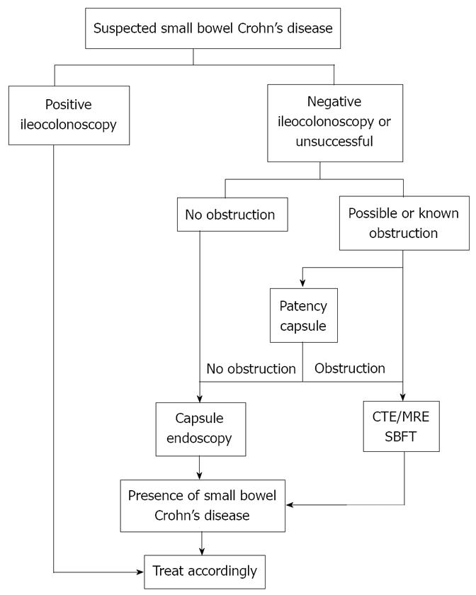
Figure 2 Algorithm for approaching suspected Crohn’s disease.
(Leighton et al[19]).
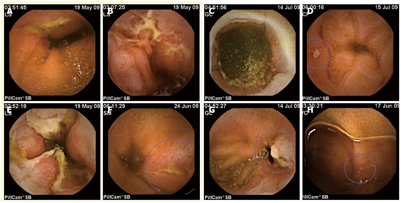
Figure 3 CE findings for Crohn’s disease are not specific.
In the images, only the ones included inside the yellow square are confirmed cases of Crohn’s disease. The other four were NSAIDs related lesions. A, B: Aftous ulcers, typical of Crohn’s disease; B, E: Geographical ulcers, observed in severe cases of small bowel Crohn’s disease, with strictures associated. D, H: Very small aftae, quite often observed in normal people, but, in these cases, in patients with a recent NSAIDs therapy. C: A typical ring-shaped stricture associated to NSAIDs. G: An ulcer in a patient with anemia and in treatment with high doses of NSAIDs for arthritis.
- Citation: Redondo-Cerezo E. Role of wireless capsule endoscopy in inflammatory bowel disease. World J Gastrointest Endosc 2010; 2(5): 179-185
- URL: https://www.wjgnet.com/1948-5190/full/v2/i5/179.htm
- DOI: https://dx.doi.org/10.4253/wjge.v2.i5.179


