©2010 Baishideng Publishing Group Co.
World J Gastrointest Endosc. Oct 16, 2010; 2(10): 349-351
Published online Oct 16, 2010. doi: 10.4253/wjge.v2.i10.349
Published online Oct 16, 2010. doi: 10.4253/wjge.v2.i10.349
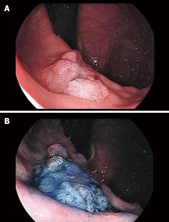
Figure 1 Endoscopic findings in the remnant stomach.
A: Conventional endoscopic observation revealed a 15 mm elevated lesion in the remnant stomach. Furthermore, it produced a viscous, mucinous secretion; B: Observation using the indigo stain showed a superficial villous design and central dip.
Figure 2 Diagnosis of the villous elevated lesion as a fistula.
A: Endoscopic ultrasonography with a 15-MHz mini-probe revealed a fistula from the central dip to the outside of the gastric wall (arrow); B: Fisterography by endoscopy showed the fistula of about 2 cm from the lesser curvature side of the remnant stomach (arrow); C: Computed tomogram just after fisterography showed the fistula between the pancreas tail and stomach (arrow).
- Citation: Uesato M, Nabeya Y, Miyazaki S, Aoki T, Akai T, Shuto K, Tanizawa T, Miyazaki M, Matsubara H. Postoperative recurrence of an IPMN of the pancreas with a fistula to the stomach. World J Gastrointest Endosc 2010; 2(10): 349-351
- URL: https://www.wjgnet.com/1948-5190/full/v2/i10/349.htm
- DOI: https://dx.doi.org/10.4253/wjge.v2.i10.349


