©2010 Baishideng.
World J Gastrointest Endosc. Jan 16, 2010; 2(1): 3-9
Published online Jan 16, 2010. doi: 10.4253/wjge.v2.i1.3
Published online Jan 16, 2010. doi: 10.4253/wjge.v2.i1.3
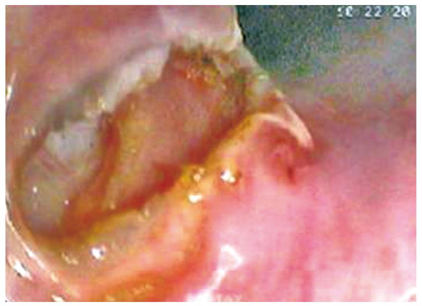
Figure 1 Endoscopic view showing access to the mediastinum following a full thickness incision with a needle knife alone.
Reproduced with permission from Fritscher-Ravens et al[23].
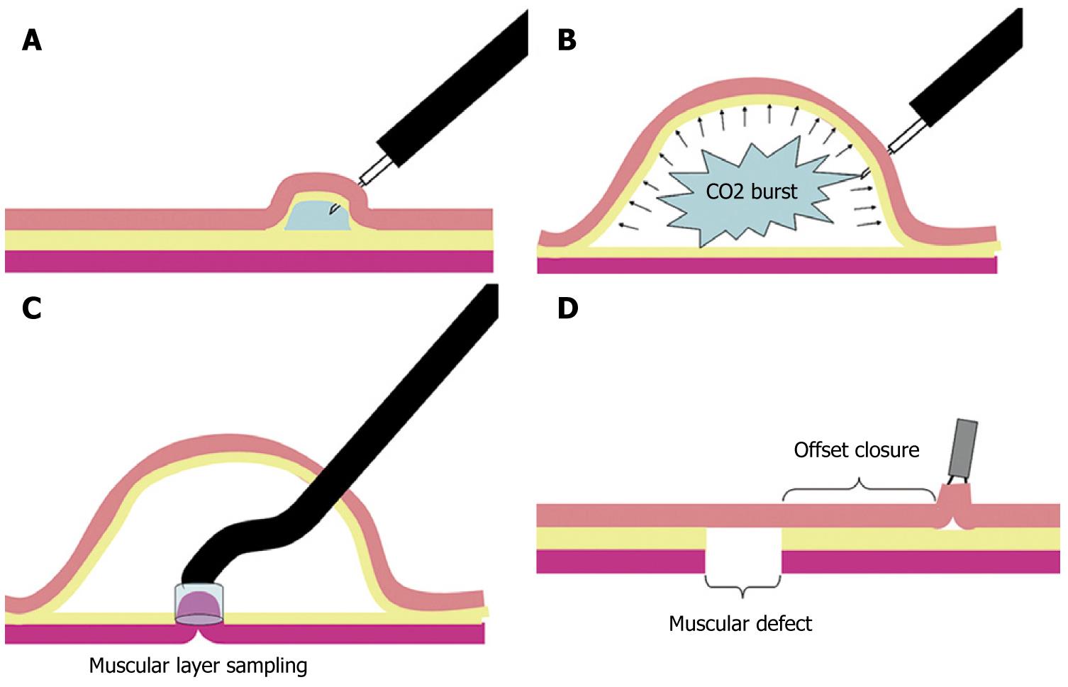
Figure 2 Transesophageal mediastinoscopy technique.
A: Saline-solution-injection test to confirm needle-tip entry into the submucosa; B: Gas submucosal dissection with high-pressure CO2; C: Muscular-layer resection with cap-EMR technique inside of the submucosal space; D: Offset closure of the muscular defect with overlying mucosal flap. Reproduced with permission from Sumiyama et al[24].
Figure 3 This figure outlines the process of transesophageal entry into the mediastinum and shows representative flexible endoscopic views.
A: Endoscopic view of the Duette Band Mucosectomy device. A band is placed around a small segment of esophageal mucosa and a snare is placed around the entrapped mucosa. Electrocautery is applied through the snare to accomplished resection of the mucosa; B: Endoscopic view of the esophageal lumen (L) and the submucosal tunnel (T); C: Endoscopic view from within the submucosal tunnel. A needle knife, pictured in the lower right corner of the image, is used to create and esophageal exit site (indicated by the black arrow); D: View of the mediastinum with the lateral esophageal wall on the left and pleura on the right; E: Endoscopic view of the lung and pleura. The black arrow shows a tear in the pleura created by biopsy forceps, which permits entry into the chest cavity; F: Endoscopic view of the chest cavity structures including the lung apex (A), lung (L), thoracic vertebra (TV), rib (R), and intercostal space (IS). Figure 3A-C and 3E are reproduced with permission from Gee et al[21] Figure 3D is reproduced with permission from Willingham et al[22].
- Citation: Turner BG, Gee DW. Natural orifice transesophageal thoracoscopic surgery: A review of the current state. World J Gastrointest Endosc 2010; 2(1): 3-9
- URL: https://www.wjgnet.com/1948-5190/full/v2/i1/3.htm
- DOI: https://dx.doi.org/10.4253/wjge.v2.i1.3


