
















Copyright: ©Author(s) 2026.
World J Gastrointest Endosc. Aug 16, 2026; 18(8): 122451
Published online Aug 16, 2026. doi: 10.4253/wjge.122451
Published online Aug 16, 2026. doi: 10.4253/wjge.122451
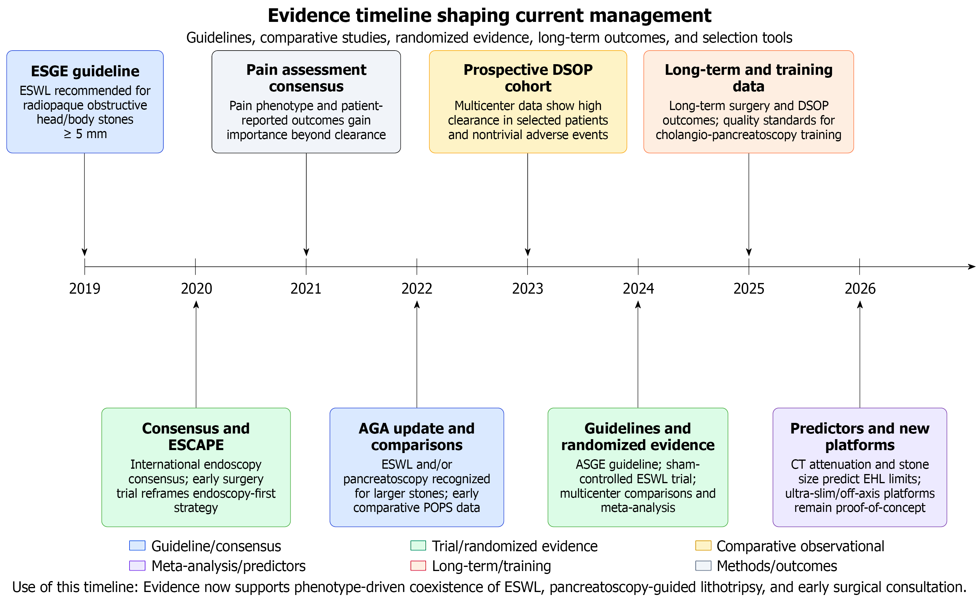
Figure 1 Evidence timeline shaping current management of difficult pancreatic duct stones.
Chronological overview of key guideline statements, randomized trials, comparative observational studies, meta-analyses, long-term outcome studies, imaging-predictor studies, and training or safety statements relevant to extracorporeal shock wave lithotripsy, pancreatectomy-guided lithotripsy, and the interface with pancreatic surgery. ESGE: European Society of Gastrointestinal Endoscopy; ESWL: Extracorporeal shock wave lithotripsy; DSOP: Digital single-operator pancreatoscopy; AGA: American Gastroenterological Association; ASGE: American Society for Gastrointestinal Endoscopy; CT: Computed tomography; EHL: Electrohydraulic lithotripsy.
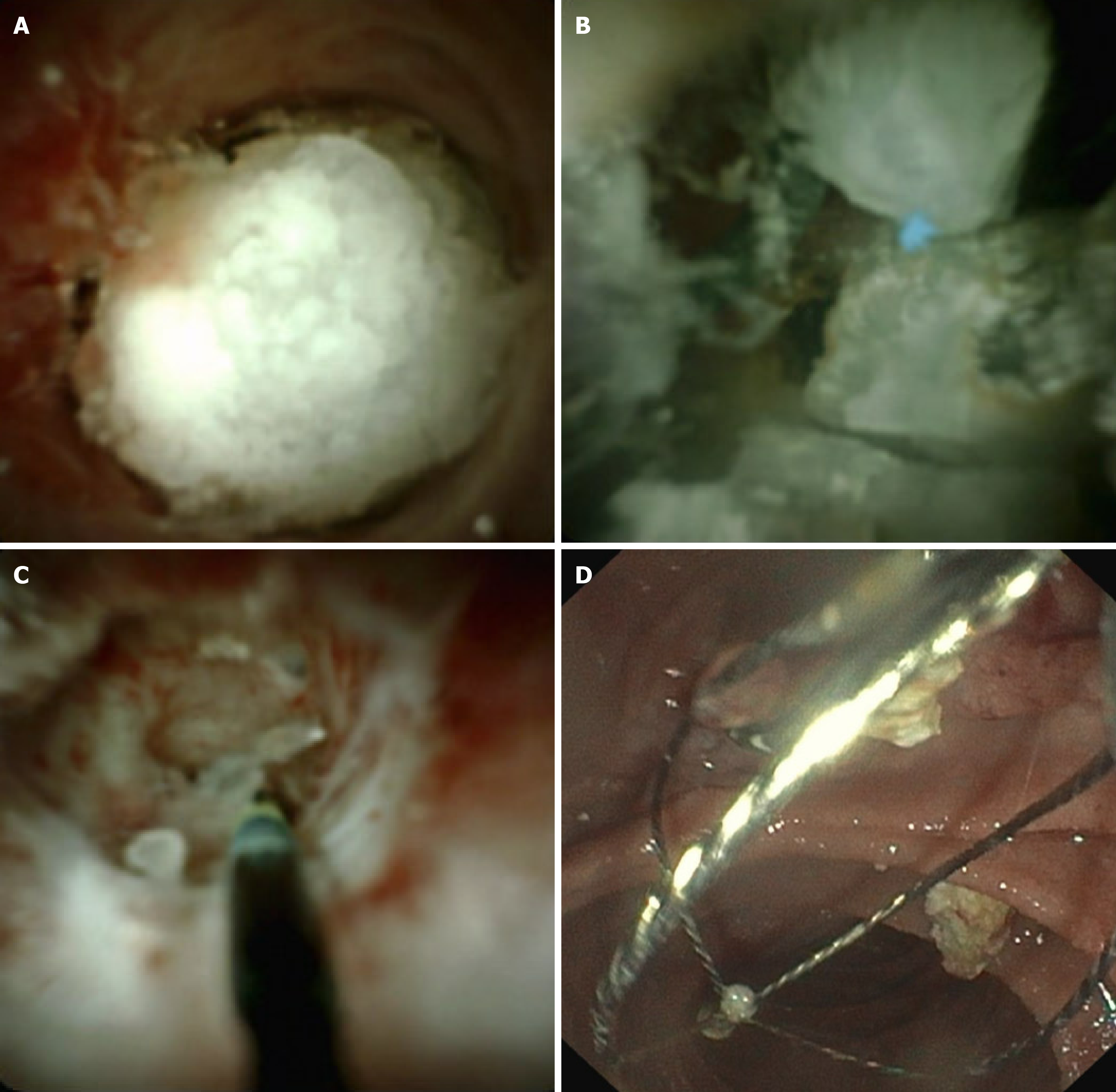
Figure 2 Representative pancreatoscopy-guided lithotripsy workflow.
A: Direct pancreatoscopy shows an obstructing pancreatic duct stone within the main pancreatic duct; B: A laser fiber or electrohydraulic lithotripsy probe is positioned against the stone under direct visualization; C: Controlled intraductal lithotripsy achieves progressive stone fragmentation; D: Residual fragments are cleared, and associated downstream ductal stricture is managed with dilation and/or temporary pancreatic stenting when required.
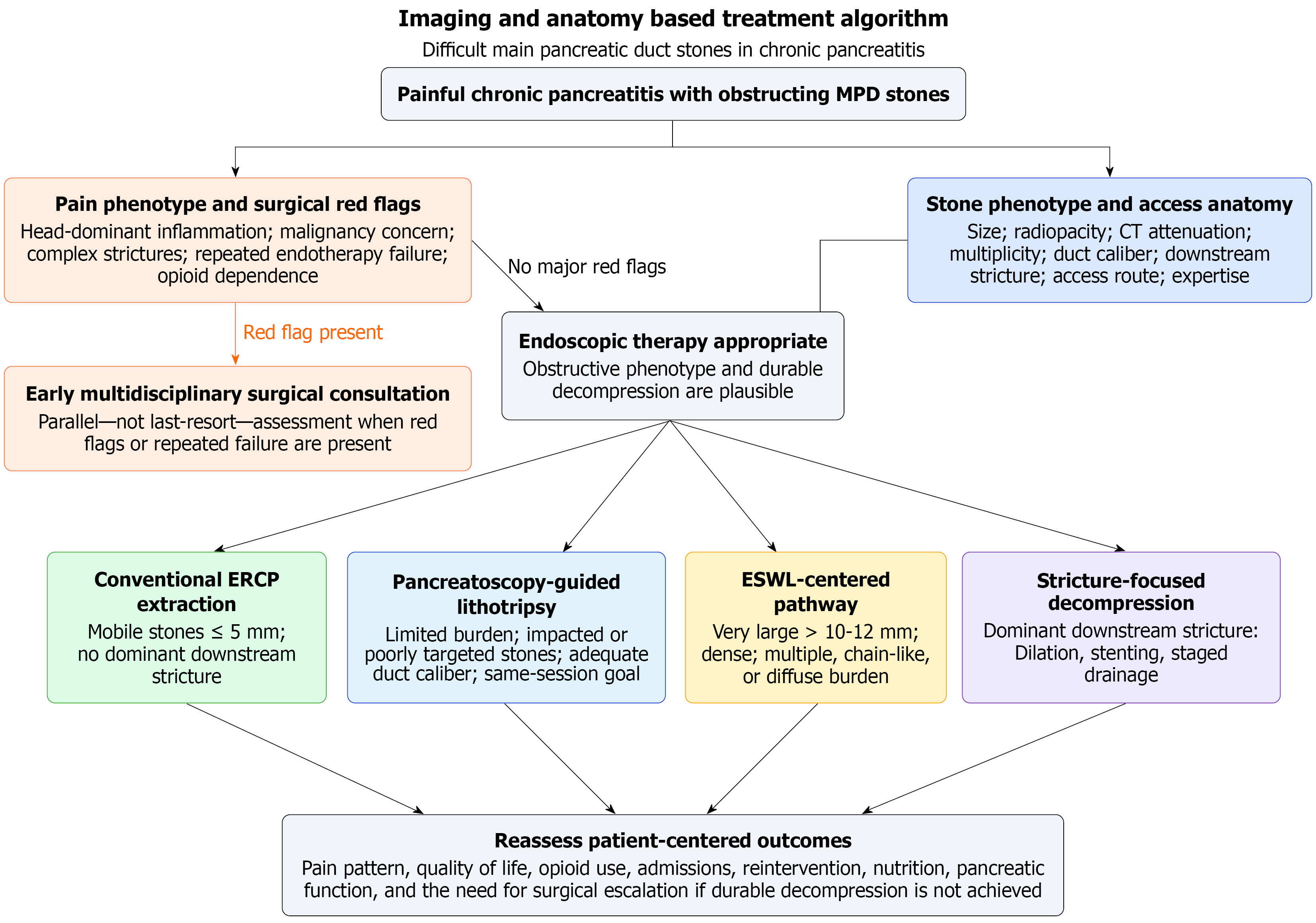
Figure 3 Phenotype-driven treatment algorithm for difficult main pancreatic duct stones.
Proposed clinical algorithm for difficult pancreatic duct stones, incorporating patient symptoms, stone and duct features, access anatomy, local expertise, and expected treatment burden to guide selection among endoscopic retrograde cholangiopancreatography extraction, pancreatoscopy-guided lithotripsy, extracorporeal shock wave lithotripsy-centered therapy, stricture-focused decompression, and early surgical consultation. MPD: Main pancreatic duct; CT: Computed tomography; ERCP: Endoscopic retrograde cholan
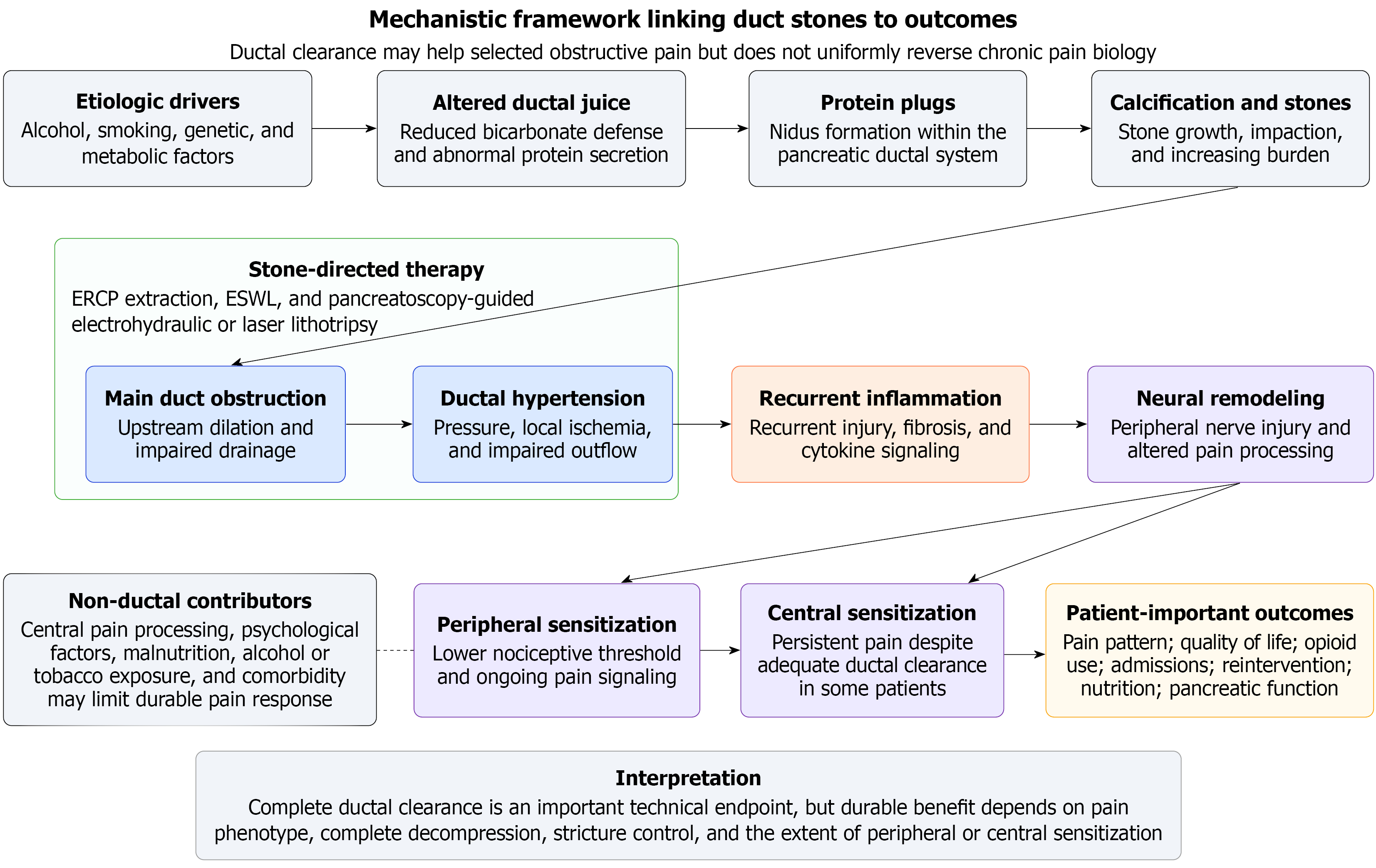
Figure 4 Mechanistic framework linking ductal stone therapy to patient-important outcomes.
Schematic overview of how protein plugs and calcification may lead to main pancreatic duct obstruction, ductal hypertension, recurrent inflammation, neural remodeling, and peripheral or central sensitization. The figure illustrates why ductal clearance can help selected patients with obstructive disease, but may not consistently improve pain, quality of life, opioid use, or the need for later surgery. ERCP: Endoscopic retrograde cholangiopancreatography; ESWL: Extracorporeal shock wave lithotripsy.
- Citation: Hashimoto R. Pancreatoscopy guided lithotripsy for difficult pancreatic duct stones: Evidence and treatment selection in chronic pancreatitis. World J Gastrointest Endosc 2026; 18(8): 122451
- URL: https://www.wjgnet.com/1948-5190/full/v18/i8/122451.htm
- DOI: https://dx.doi.org/10.4253/wjge.122451