
















Copyright: ©Author(s) 2026.
World J Gastrointest Endosc. Jul 16, 2026; 18(7): 123520
Published online Jul 16, 2026. doi: 10.4253/wjge.123520
Published online Jul 16, 2026. doi: 10.4253/wjge.123520
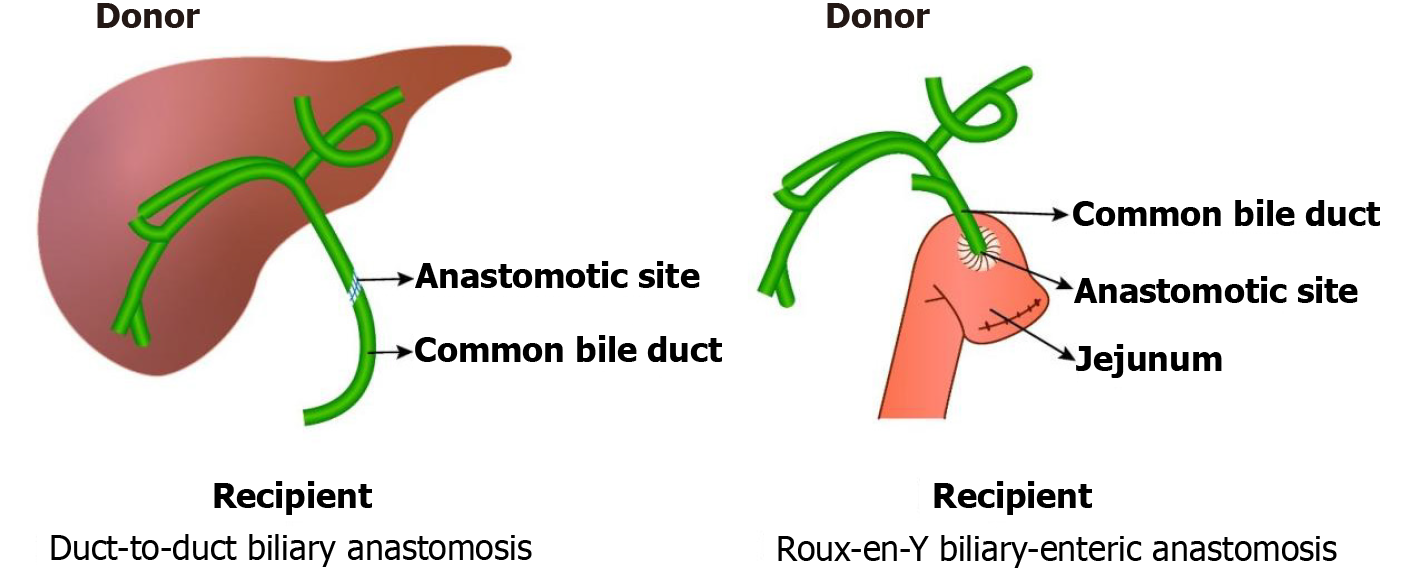
Figure 1
Duct-to-duct biliary anastomosis and Roux-en-Y biliary-enteric anastomosis.
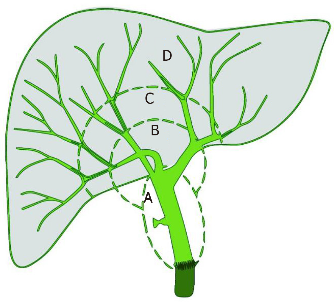
Figure 2 Four anatomical zones of biliary tree of non-anastomotic biliary stricture classified by Buis classification.
Zone A: Hilar bifurcation; zone B: Central intrahepatic ducts; zone C: Peripheral intrahepatic ducts; zone D: Terminal/septal ducts.
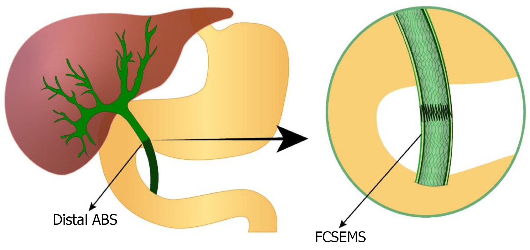
Figure 3 Endoscopic therapy of distal anastomotic biliary stricture with fully covered self-expandable metal stents.
ABS: Anastomotic biliary stricture; FCSEMS: Fully covered self-expandable metal stents.
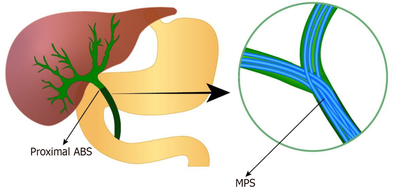
Figure 4 Endoscopic therapy of proximal anastomotic biliary stricture with multiple plastic stents.
ABS: Anastomotic biliary stricture; MPS: Multiple plastic stents.
- Citation: Shrestha UK. Mitigating challenges in endoscopic management of post-liver transplant biliary strictures. World J Gastrointest Endosc 2026; 18(7): 123520
- URL: https://www.wjgnet.com/1948-5190/full/v18/i7/123520.htm
- DOI: https://dx.doi.org/10.4253/wjge.123520