















Copyright: ©Author(s) 2026.
World J Gastrointest Endosc. Jul 16, 2026; 18(7): 121212
Published online Jul 16, 2026. doi: 10.4253/wjge.121212
Published online Jul 16, 2026. doi: 10.4253/wjge.121212
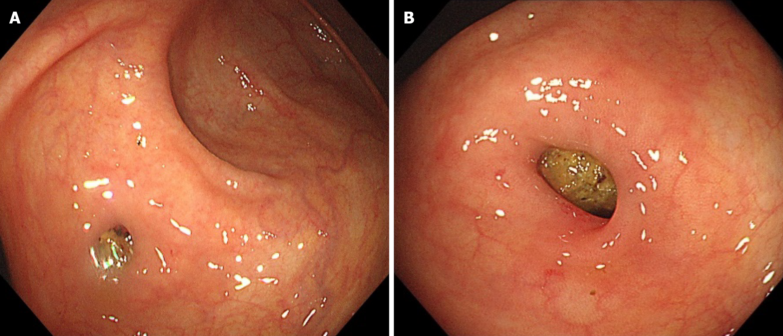
Figure 1 Endoscopic view.
A: Orifice of vermiform appendix; B: Fecal stones in the appendix.
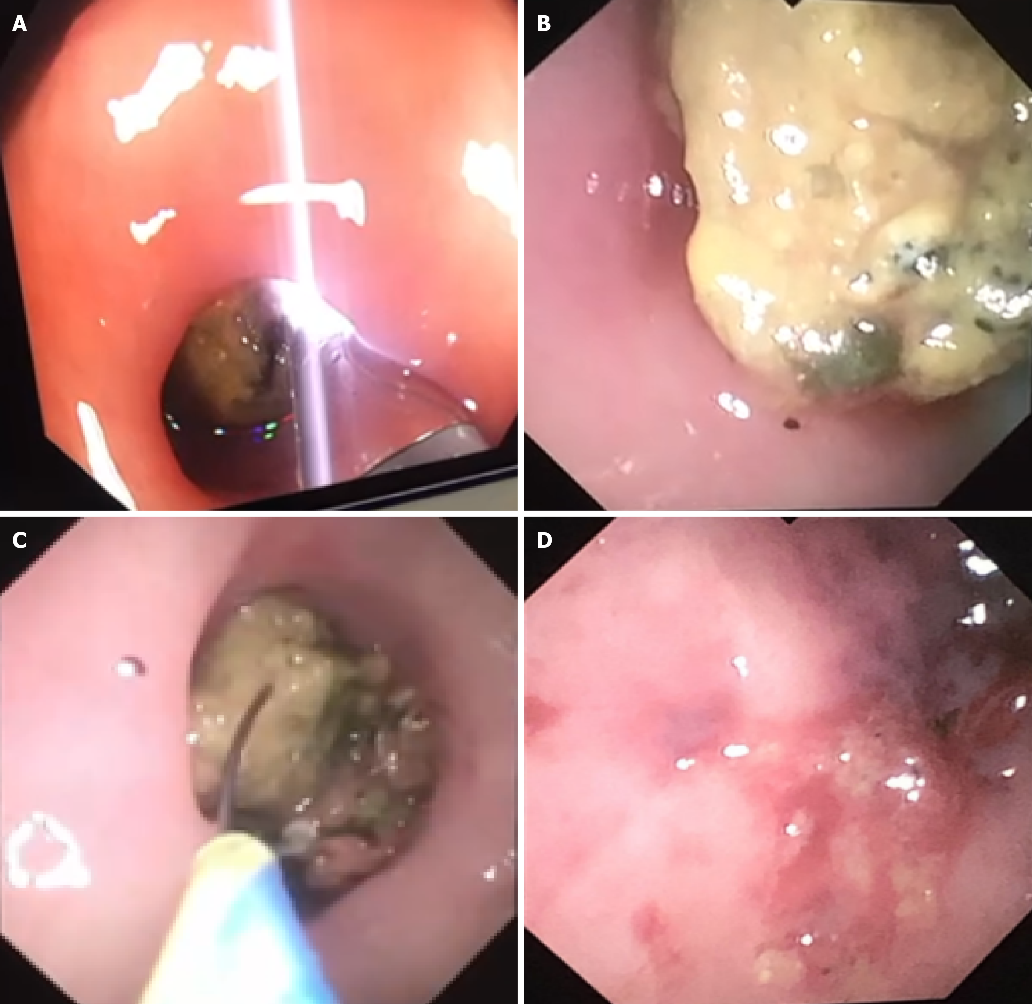
Figure 2 EyeMAX view.
A: Insertion of eyeMAX; B: Observation of fecal stones in the stump appendix; C: Stone retrieval basket through eyeMAX into stump appendix; D: Edema of the inner wall of the stump appendix after fecal stone was dislodged.
- Citation: Li W, Xiao M, Wu DD, Zhang KG, Ye C. Stump appendicolith removal using a pancreaticobiliary scope in an elderly patient: A case report. World J Gastrointest Endosc 2026; 18(7): 121212
- URL: https://www.wjgnet.com/1948-5190/full/v18/i7/121212.htm
- DOI: https://dx.doi.org/10.4253/wjge.121212