
















Copyright: ©Author(s) 2026.
World J Gastrointest Endosc. Jul 16, 2026; 18(7): 119987
Published online Jul 16, 2026. doi: 10.4253/wjge.119987
Published online Jul 16, 2026. doi: 10.4253/wjge.119987
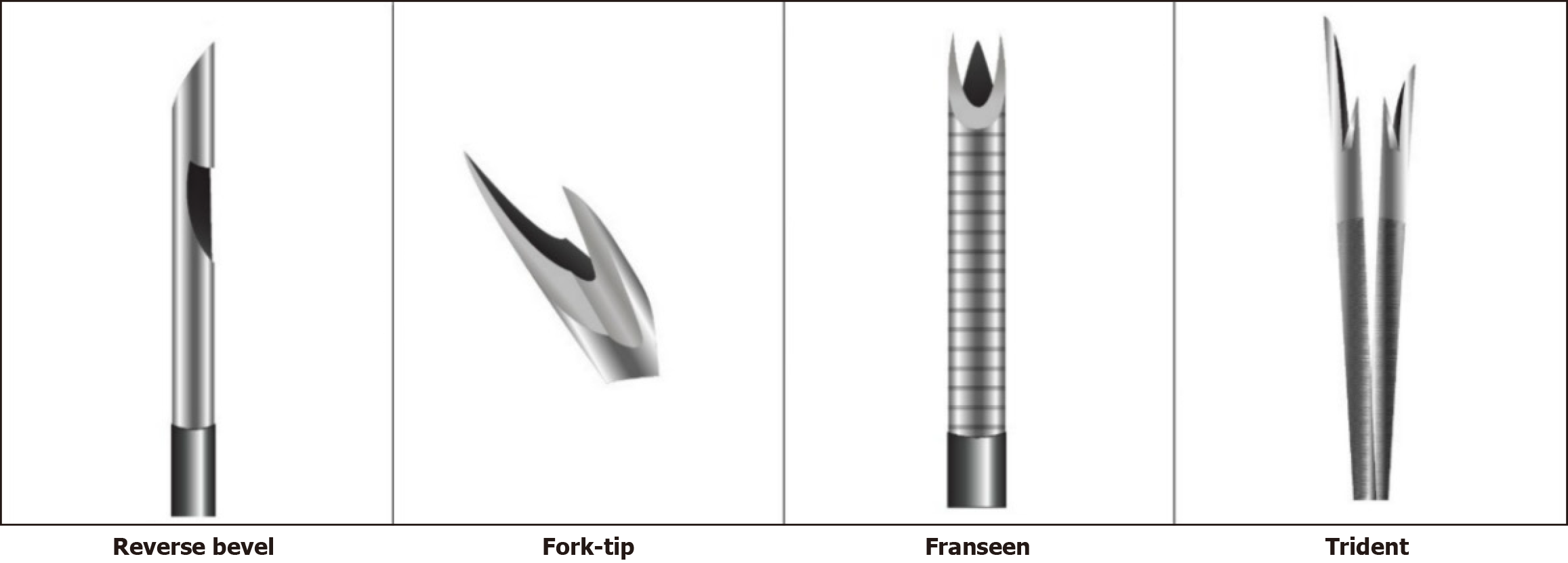
Figure 1 Fine-needle biopsy needles for endoscopic ultrasound-guided tissue acquisition.
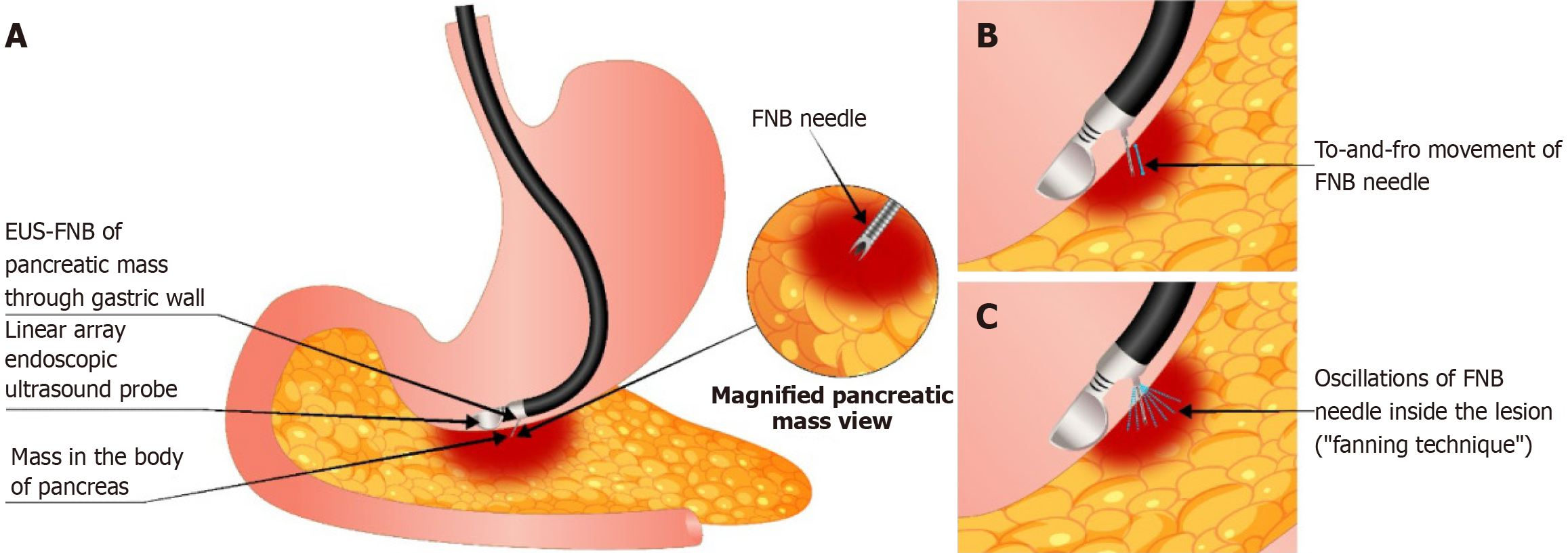
Figure 2 Endoscopic ultrasound-guided fine-needle biopsy of a mass.
A: Transgastric endoscopic ultrasound-guided fine-needle biopsy of a mass in the body of the pancreas; B: To-and-fro movement of the Fine-needle biopsy needle; C: Oscillations of the Fine-needle biopsy needle inside the lesion (“fanning technique”). EUS-FNB: Endoscopic ultrasound-guided fine-needle biopsy; FNB: Fine-needle biopsy.
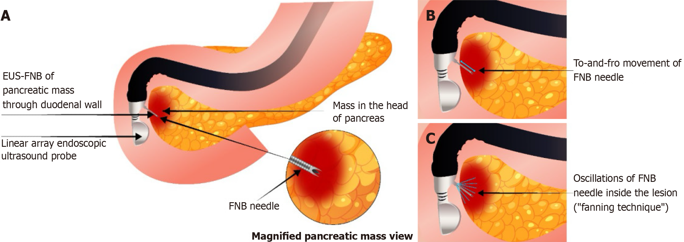
Figure 3 Transgastric endoscopic ultrasound-guided.
A: Transduodenal endoscopic ultrasound-guided fine-needle biopsy of a mass in the head of the pancreas; B: To-and-fro movement of the fine-needle biopsy needle; C: Oscillations of the fine-needle biopsy needle inside the lesion (“fanning technique”). EUS-FNB: Endoscopic ultrasound-guided fine-needle biopsy; FNB: Fine-needle biopsy.
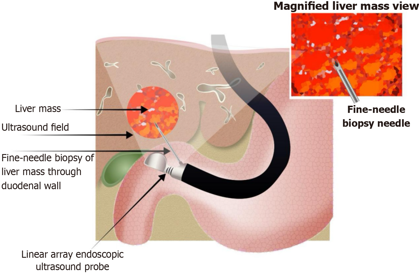
Figure 4 Endoscopic ultrasound-guided fine-needle biopsy of liver mass.
- Citation: Shrestha UK. Preventive strategies for complications after endoscopic ultrasound-guided fine-needle biopsy. World J Gastrointest Endosc 2026; 18(7): 119987
- URL: https://www.wjgnet.com/1948-5190/full/v18/i7/119987.htm
- DOI: https://dx.doi.org/10.4253/wjge.119987